|
The Effects of Selected Pyrethroids on Embryos of Bombina bombina during different Culture and Semi-field Conditions 3 Results
Egg deposition of Bombina bombina occurred 9-12 h after injection of human chorionic gonadotropin. About 75-400 eggs were found in the breeding aquarium, on the aquatic vegetation and plastic plants as well as on the small sticks. 3.1 Embryo development in control groups at different culture conditionOnly the best-fertilised eggs from a given breeding aquarium were used and only normally cleaving embryos were used in the tests. Control mortality could be kept to less than 5% when this procedure was followed. 3.1.1 Embryo development in FETAX solutions at two different temperaturesControls At 24°C, the control group of Bombina bombina reached stage 46 after 120 h. A stage 46 larvae were recognised by the appearance of the hind limb bud, the coiling of the gut, and the shape of the operculum covering the gills. The best indicator that the larvae had attained at stage 46 was the appearance of the hind limb bud. Gut coiling was also easily observed at stage 46 (at stage 45 embryos do not display complete tight gut coiling). At stage 46, the larvae were about 9.5 mm in length. Embryos of Bombina bombina, kept at 24°C, were still in the jelly coat after 24 h and were not free until after about 72 h. At 20°C, the control group reached stage 46 according to Nieuwkoop and Faber (1975) after 216 h. At stage 46, the larvae were about 9.5 mm in length. Most of embryos, kept at 20°C, were still in the jelly coat after 96 h. After 120 h most of the embryos were free, however, a few were not free until after 144 h. The development of the control groups of Bombina bombina at the two different temperatures is illustrated in table 3.1. For comparison with the development of Xenopus normally used for the standard FETAX test, selected staging according to Nieuwkoop and Faber (1975) is also included. Table 3.1 Effekten af temperaturen på udviklingen af Bombina bombina embryoer. Embryostadiet blev bestemt ved hjælp af Gosner (1960).
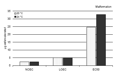
* Stage according to Nieuwkoop and Faber (1975) for comparison with Xenopus. 3.1.2 Embryo development in two different mediaControls The results revealed that the embryo development was identical in the two media, FETAX and pond water. After 216 h, the control group reached stage 46 in both media at 20°C and all embryos were normal and showed no malformations. At stage 46, the larvae were about 9.5 mm in length in both media. 3.2 Tests with reference substanceEffect of 6-aminonicotinamide The results revealed that the effect of 6-aminonicotinamide in both pond water and the FETAX solutions with Bombina bombina were comparable with the effect described for Xenopus in the ASTM standards. The EC50 value for malformations after 120 h and 216 h at 24°C and 20°C, respectively, was 4.9 mg/l in FETAX (at both temperatures) and 5.2 mg/l in pond water. The LC50 value was about 25000 mg/l in FETAX and 26000 in pond water (mean values of 9 experiments; SD of the mean less than 10%). The results revealed that 55% and 58% of the embryos had malformation at 5.5 mg/l 6-aminonicotinamide at 24°C and 20°C, respectively. A mortality of 44% and 51% was found at a concentration of 2500 mg/l aminonicotinamide at 24°C and 20°C, respectively. The most conspicuous malformations caused by 6-aminonicotinamide were severe optic cup rupture, edema and facial malformations such as ocular edema and foreshortened facial features. In addition, severe heart malformations were also seen. 3.3 Effects of esfenvalerate on Bombina bombina at two different temperaturesSince the renewal procedure entailed fresh replacement of the test material every 24 h during the test and the chemical measurement from previous experiments did not reveal a decrease of the pesticide concentration during the exposure period (Larsen and Sørensen, 2004), the results of the present study are expressed as nominal concentrations. 3.3.1 In vivo observationsEsfenvalerate Esfenvalerate affected the embryos gradually during the exposure in a dose depended manner at both temperatures. After 24 h, no effect of esfenvalerate was seen on the embryos even at the highest concentration compared with the controls at both temperatures. After 48 h, no effect of esfenvalerate was seen on the embryos even at the highest concentration compared with the controls at 20°C. At 24°C, no effect was seen at concentrations up to 5 μg/l, however, at 5 g/l beginning twisting of the embryos was seen now and then. The spasmodic twisting increased in intensity with increasing concentrations of esfenvalerate and at 150 and 300μg/l, the embryos had constant spasmodic twisting. At concentrations from 50 μg/l, the spasmodic twisting of the embryos caused that some of the embryos had already left the jelly coat at this stage. In the control group, only a few embryos had begun to leave the jelly coat after 48 h. At 100, 150, and 300 μg/l, edema was seen in some of the embryos. After 72 h, no effect of esfenvalerate was seen on the embryos at 20°C even at the highest concentration compared with the controls, and all embryos were in the jelly coat. At 24°C, several of the embryos in the control group had begun to leave the jelly coat and all the embryos exposed to esfenvalerate concentrations above 50 μg/l were free. Periodic spasmodic twisting was seen in most of the embryos above 5 μg/l and in 50 μg/l constant spasmodic twisting was seen. At concentrations above 100 μg/l, occasionally blistering and cardiac edema appeared. In 150 and 300 μg/l, blistering and cardiac edema were frequently seen. The embryos were immobilised by constant spasmodic twisting. After 96 h, the first effects of esfenvalerate were seen at 20°C. No effect was seen at concentrations up to 5 μg/l, however, at 5 μg/l beginning twisting of the embryos was seen now and then. The spasmodic twisting increased in intensity with increasing concentrations of esfenvalerate and at 300 μg/l the embryos had constant spasmodic twisting. At concentrations from 10 μg/l, the spasmodic twisting of the embryos caused that some of the embryos had left the jelly coat at this stage. In the control group, only a few embryos had begun to leave the jelly coat after 96 h. At 100 μg/l, most of the embryos had left the jelly coat. At 24°C all embryos in the control group were free from the jelly coat and beginning twisting was seen at 1 μg/l. The intensity of these spasmodic twisting increased when the concentrations of esfenvalerate were increased. At concentrations above 50 μg/l, the embryos were immobilised caused by constant spasmodic twisting and occasionally blistering and cardiac edema appeared. At concentrations above 100 μg/l, blistering and edema were frequently seen as well as head and brain malformations. The heartbeat was very slow and severe heart malformations were normally seen. After 120 h, several of the embryos in the control group began to leave the jelly coat at 20°C, and all the embryos exposed to esfenvalerate concentrations above 10 μg/l were free. Periodic spasmodic twisting was seen in most of the embryos above 2.5 μg/l and in 50 μg/l constant spasmodic twisting was seen. At concentrations above 100 μg/l, occasionally blistering and cardiac edema appeared. In 150 and 300 μg/l, blistering and cardiac edema were frequently seen. The embryos were immobilised by constant spasmodic twisting. At 24°C, spasmodic twisting was seen at 1 μg/l. At concentrations above 50 μg/l, the embryos were immobilised caused by constant spasmodic twisting and blistering and edema were often seen. At concentrations above 100 μg/l, blistering and edema were frequently seen as well as head and brain malformations. The heartbeat was very slow and severe heart malformations were seen at 100, 150 and 300 μg/l. At this point the embryos had reached stage 46 and were killed and fixed as previously described. After 144, 168, and 192 the same picture was seen at 20°C. All embryos in the control group were free from the jelly coat and beginning twisting was seen at 2.5 μg/l. The intensity of these spasmodic twisting increased when the concentrations of esfenvalerate were increased. At concentrations above 50 μg/l, the embryos were immobilised caused by constant spasmodic twisting and occasionally blistering and cardiac edema appeared. At concentrations above 100 μg/l, blistering and edema were frequently seen as well as head and brain malformations. The heartbeat was very slow and severe heart malformations were normally seen. After 216 h, spasmodic twisting was seen at 1 μg/l. At concentrations above 50 μg/l, the embryos were immobilised caused by constant spasmodic twisting and blistering and edema were often seen. At concentrations above 100 μg/l, blistering and edema were frequently seen as well as head and brain malformations. The heartbeat was very slow and severe heart malformations were seen at 100, 150 and 300 μg/l. At this point the embryos had reached stage 46 and were killed and fixed as previously described. 3.3.2 Recordings of malformation, growth inhibition and mortality at the end of the test (120 h and 216 h for 24°C and 20°C, respectively)Malformation Most abnormal embryos possessed multiple malformations. However, at 1 and 2.5 μg/l no malformations were seen and the larvae were at the normal stage 46 according to Nieuwkoop and Faber (1975) and comparable with the controls in all respects. Above a concentration of 5 μg/l, gut abnormalities were often seen indicated by a loose gut coiling. At 10 μg/l in addition to gut abnormalities, a relatively mild flexure of the tail was normally seen. Embryos with a sloping forehead and distended mouth were often seen. The head and face region of these embryos was malformed. The face was flattened and the forebrain was deflected downward along the front of the brow. Heart malformations were also observed at concentrations above 10 μg/l. Increasing concentrations of esfenvalerate resulted in increasingly severe heart malformations. Often the heart showed an abnormal expansion of the ventricle just in front of the gut. At 50 μg/l, blistering and edema were common and various degrees of undeveloped gills were also seen. At 24°C, about 20% of the embryos had malformations at 10 μg/l, and more than 75% of the embryos exposed to 100 μg/l and higher concentrations had malformations. At 300 μg/l, all embryos had malformations. At 20°C, about 30% of the embryos had malformations at 10 μg/l and about 90% of the embryos exposed to 100 μg/l and higher concentrations had malformations. At 300 μg/l all embryos had malformations. The EC50 value for malformations was calculated to be 32.7 and 24.7 μg/l for 24°C and 20°C, respectively (further details cf. Appendix C). The most significant malformation observed included cardiac malformations, severe lateral flexure, edema, notochord, brain and gut malformations. Effect concentrations are shown in figure 3.1. Growth inhibition After 120 h and 216 h (for 24°C and 20°C, respectively) of exposure to concentrations up to 300 g/l, the mean length of the embryos was at both temperatures comparable with the length of the control group. The mean length of embryos in the control group was 9.5 mm (S.D. 0.08) and 9.3 mm. (S.D. 0.3), in 24°C and 20°C, respectively. The NOEC, LOEC, and LC50 values for growth inhibition were all higher than 300 μg/l. Mortality No significant mortality was seen at concentrations up to 300 μg/l at both temperatures. The NOEC, LOEC, and LC50 values were all higher than 300 μg/l. Teratogen index (TI) The effects of esfenvalerate on TI on Bombina bombina at different temperatures are depicted in table 3.2. Table 3.2 Teratogen index (TI) for esfenvalerat ved to forskellige temperaturer.
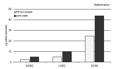
Figure 3.1 En sammenligning af misdannelserne forårsaget af esfenvalerat på Bombina bombina ved to forskellige temperaturer. Resultaterne er udtrykt ved angivelse af NOEC, LOEC og EC50/LC50 værdierne efter 120 og 216 timers eksponering ved henholdsvis 24 og 20°C. Efter denne eksponeringstid var embryonerne i de respektive kontrolgrupper nået til stadie 46. Resultaterne er gennemsnit af 3 forsøg. Standardafvigelsen af middeltallene var mindre end 10%. 3.4 Effects of esfenvalerate on Bombina bombina in different culture mediaThe effect of esfenvalerate on malformation, growth inhibition, and mortality in Bombina bombina was tested in pond water and the effects were compared with that found in the FETAX solution. After 216 h of exposure at 20°C, the embryos in the control group were at stage 46 corresponding to the stage of the controls at the end of the test in the standard FETAX test with Xenopus in both media. 3.4.1 In vivo observationsEsfenvalerate Esfenvalerate affected the embryos gradually during the 216 h exposure in a dose depended manner in both media. The same effects of esfenvalerate were seen in both media; however, the effects were more distinct in the FETAX solution. No effect of esfenvalerate was seen on the embryos even at the highest concentration compared with the controls during the first 72 h of exposure. After 96 h, the first effects of esfenvalerate were seen in the FETAX medium at 5 μg/l where beginning twisting of the embryos was seen now and then. The spasmodic twisting increased in intensity with increasing concentrations of esfenvalerate and at 300 μg/l the embryos had constant spasmodic twisting. At concentrations from 10 μg/l some of the embryos had left the jelly coat and at 100 μg/l most of the embryos had left the jelly coat. The same pattern was seen in pond water, however, in this medium no effect was seen at concentrations up to 10 μg/l. In pond water beginning twisting was first seen at 50 μg/l. At concentrations from 50 μg/l some of the embryos had left the jelly coat. In the control group, only a few embryos had left the jelly coat after 96 h in both media. After 120 h, several of the embryos in the control group began to leave the jelly coat in both media, and all the embryos exposed to esfenvalerate concentrations above 10 μg/l were free. Periodic spasmodic twisting was seen in most of the embryos above 2.5 μg/l and 10 μg/l in FETAX and pond water, respectively. At concentrations above 100 μg/l spasmodic twisting was significant and occasionally blistering and cardiac edema appeared in both media. In 150 and 300 μg/l, blistering and cardiac edema were frequently seen. The embryos were immobilised by constant spasmodic twisting in both media at concentrations of 150 and 300 μg/l. After 144, 168, and 192 the same picture was seen in both media. All embryos in the control group were free from the jelly coat. The intensity of the spasmodic twisting increased with increasing concentrations of esfenvalerate. Periodic spasmodic twisting was seen in most of the embryos at concentrations above 50 μg/l in the pond water. In FETAX, the embryos were immobilised caused by constant spasmodic twisting at these concentrations and occasionally blistering and cardiac edema appeared. At concentrations above 100 μg/l, blistering and edema were frequently seen in both media as well as head and brain malformations. The heartbeat was very slow and severe heart malformations were normally seen. After 216 h, spasmodic twisting was seen at 1 μg/l and 5 μg/l in FETAX and pond water, respectively. At concentrations above 50 μg/l and above 100μg/l, in FETAX and pond water, respectively, the embryos were immobilised caused by constant spasmodic twisting as well as blistering and edema were often seen. At concentrations above 100 μg/l, blistering and edema were frequently seen in both media as well as head and brain malformations. Furthermore, the heartbeat was very slow and severe heart malformations were seen at 100, 150 and 300 μg/l. 3.4.2 Recordings of malformation, growth inhibition, and mortality at the end of the testMalformation Most abnormal embryos possessed multiple malformations. However, at 1 and 2.5 μg/l no malformations were seen and the larvae were at the normal stage 46 according to Nieuwkoop and Faber (1975) and comparable with the controls in all respects. At concentrations above 5 μg/l, gut abnormalities were often seen in FETAX indicated by a loose gut coiling, however, no significant effects was found in embryos in pond water. At 10 μg/l in addition to gut abnormalities, a relatively mild flexure of the tail was normally seen. Embryos with a sloping forehead and distended mouth were often seen. The head and face region of these embryos was malformed. The face was flattened and the forebrain was deflected downward along the front of the brow. Heart malformations were also observed at concentrations above 10 g/l. Increasing concentrations of esfenvalerate resulted in increasingly severe heart malformations. The heart often showed an abnormal expansion of the ventricle just in front of the gut. At 50 μg/l, blistering and edema were common and various degrees of undeveloped gills were seen, too. In FETAX solution about 30% of the embryos had malformations at 10 μg/l and about 90% of the embryos exposed to 100 μg/l and higher concentrations had malformations. In pond water a significant effect of esfenvalerate was seen at 50 μg/l where about 15% of the embryos had malformations. In pond water at 100 μg/l and at 300 μg/l nearly all embryos had malformations in both media. The EC50 value for malformations was calculated to be 24.7 and 43.7 μg/l in FETAX solution and pond water, respectively (Further detail cf. Appendix C). The most significant malformation observed included cardiac, severe lateral flexure, edema, notochord, brain, and gut malformations. Effect concentrations for malformation are shown in figure 3.2 Growth inhibition After 216 h of exposure to concentrations up to 300 μg/l, the mean length of the embryos was in both media comparable with the length of the control group. The mean length of embryos in the control group was 9.3 mm. (S.D. 0.3) and 9.5 mm (S.D. 0.2) in FETAX and pond water, respectively. Thus, the results revealed that for growth inhibition NOEC, LOEC and the EC50 valued were higher than 300 μg/l in both media. Mortality No significant mortality was seen at concentrations up to 300 μg/l in both media. The NOEC, LOEC, and LC50 values were all higher than 300 μg/l. Teratogen index (TI) The effects of esfenvalerate on IT on Bombina bombina in different media were comparable. The TI was >12.1 and > 6.9 in FETAX and pond water, respectively.
Figure 3.2 En sammenligning af effekterne af esfenvalerat på Bombina bombina i to forskellige medier udtrykt ved sublethale misdannelser. Resultaterne er udtrykt ved angivelse af NOEC, LOEC og EC50 værdierne efter 216 timers eksponering i henholdsvis FETAX opløsning og i damvand. Efter denne eksponeringstid var embryoerne i de respektive kontrolgrupper nået til stadie 46. Resultaterne er et gennemsnit af 3 forsøg. Standardafvigelsen af middeltallene var mindre end 10%. 3.5 Recovery experiments using embryos of Bombina bombina3.5.1 The effects of esfenvalerate on Bombina bombina during the toxicity testControls During the 216 h toxicity assay the embryo development of the control group was normal. After 216 h, the embryos were at stage 46, and the head-tail length was 9.0 mm (S.D. = 0.1). Malformation was seen in 5.9% (S.D. = 1.7) of the surviving embryos. The mortality was 9.3%, which corresponds to the normal range in these tests. Embryos exposed to 20 μg/l esfenvalerate No effect of esfenvalerate was seen on the embryos during the first 96 h. After 120 h all embryos were free from the jelly coat and periodic spasmodic twisting was seen. The intensity of these spasmodic twisting was increased with increasing time of exposure. Malformations were seen in 38.9% (S.D. = 4.1) of the embryos at the end of the exposure. Most abnormal embryos possessed multiple malformations including gut abnormalities as loose gut coiling, mild flexure of the tail, sloping forehead, and heart malformation. At the end of the test, a mortality of 7.3% was found and the surviving embryos were at stage 46 with a length of 9.2 (S.D. = 0.2) mm. Embryos exposed to 125 μg/l esfenvalerate During the first 72 h of exposure no effect was seen; however, after 96 h, the first effects of esfenvalerate were seen. These effects were spasmodic twisting and most of the embryos had left the jelly coat at this stage. After 120 h, constant spasmodic twisting was seen in many embryos, furthermore blistering and cardiac edema were frequently seen. After 144-216 h, all embryos were immobilised caused by constant spasmodic twisting. Blisting and edema were frequently seen as well as gut abnormalities, flexure of the tail, head and brain malformations. 83.2% (S.D. = 0.2) of the embryos had malformations at the end of the exposure. The mortality was 6% and the mean length of the surviving embryos was 8.8 mm (S.D. = 0.2) at the end of the exposure. 3.5.2 Embryo and larval development during the first part of the recovery periodImmediately after transferring the embryos to fresh medium without pesticides most of the embryos in the control group started to eat algae and moved around searching for food. About 70% of the embryos previously exposed to 20 μg/l esfenvalerate gradually recovered and were comparable with the controls. 24 h after the transfer to fresh medium, however, about 30% of this group did not ingest algae and spasmodic twisting was seen now and then. After 24 h about 80% of the embryos previously exposed to 125 μg/l esfenvalerate had still spasmodic twisting and the heartbeat of these embryos was very slow; however, about 20% of the embryos in this group were swimming around now and then. Two days after transferring the embryos to fresh medium all living larvae were swimming around and eating algae revealed by the green content in the gut. During the following weeks there was no difference in larval development in the three groups. After three weeks the larvae were at stage 38-45 and hind limps were developing. Five weeks after the transfer to fresh medium free hind limps were seen in all larvae and the hind limps were about one-third to half the length of the tail. No significant difference in development was found in the three different groups. The fore limps were not free even though they were pronounced. The larvae started to develop darker drawings on the skin than normal. Growth inhibition and mortality The effects of a short exposure to esfenvalerate on growth were not significant. All embryos were about 9 mm in length when they were transferred to the pesticide free medium, and all the larvae had grown to about 20 and 30 mm in length after two and three weeks, respectively. All larvae grew quickly and reached a maximum length within the metamorphosis of about 50 mm after about 5 weeks (after the transfer to fresh medium with food). The variation of the length was significantly within each group, however, not among the three groups (controls, previously exposed to 20 μg/l and 125 μg/l esfenvalerate, respectively). Thus, after five weeks the mean length (head-tail length) of the control group in the first experiment was 48 mm (S.D. 4.7), however, the smallest larvae in this group were 41 mm and the largest 58 mm. The length of larvae previously exposed to 20 μg/l esfenvalerate was in average 49 mm (S.D. 4.0) varying from 42-58 mm, and 49 mm (S.D. 5.3), varying from 42-58 mm for larvae which were previously exposed to 125 μg/l. In the second experiment the same pattern of variation was seen. In the two independent experiments the mean head-tail length after five weeks was 49.6 mm (S.D. 1.6), for the control group, 49.4 mm (S.D. 0.2) and 49.6 mm (S.D. 0.6) for larvae previously exposed for 20 and 125 μg/l esfenvalerate, respectively.
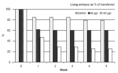
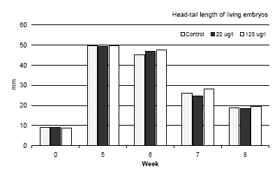
Figure 3.3 Dødeligheden af Bombina bombina i løbet af de første fem uger efter at embryonerne er blevet overført til et frisk medium uden esfenvalerat. Resultaterne er udtrykt i procent af levende embryoner, der er overført til et frisk medium (uge 0). Ved uge 0 er alle embryonerne på stadie 46. Resultaterne er gennemsnittet af to eksperimenter. Standardafvigelsen af gennemsnitsværdien var mindre end 10%. The results revealed that a high mortality was found during the first week after the larvae were transferred to the pesticide free medium and a concentration-response was found (cf. figure 3.3). Even in the control group a mortality of about 23% was found. Furthermore, it was also seen that after the first week no further mortality was found during the following period until the metamorphosis. 3.5.3 Development before and during the metamorphosis of Bombina bombinaIn vivo observations During the beginning of metamorphosis (week 6) the forelimbs became free and the notochord began to degenerate posteriorly but still normal in appearance anteriorly. At this stage the head-tail length was about 45 mm and no significant differences were seen between the three groups. The hind limb had the same length as the tail and about 60% of the larvae had free forelimbs at this stage. However, the forelimbs seemed to be bent in over the body in some of the larvae previously exposed to 20 μg/l. In this case the amphibians were not using the forelimbs and were tumbling around when they were trying to jump. All larvae previously exposed to 125 μg/l had problems with the forelimbs, which were bent in over the body and could not be used. The degradation of the tail proceeded rapidly so that the basis of the tail was affected too and after seven weeks the head-tail length was about 25 mm in all amphibians (the tail was only about 3-5 mm). A comparison of the head-tail length of Bombina bombina during metamorphosis is shown in figure 3.4. At this stage all amphibians had forelimbs; however, 50% of the amphibians previously exposed to 20 μg/l had defect forelimbs and all the amphibians previously exposed to 125 μg/l had defect forelimbs. No problems were identified in the control group. The major problem for amphibians with defect forelimbs was that they tumbled around when trying to jump on land. At the end of week 7 some of the amphibians in the control had no longer any tail and they were jumping around on land eating flies. After eight weeks all frogs in the control group had no tail and were jumping around eating flies. Amphibians previously exposed to 20 μg/l had no tail and about 20% of these animals were also jumping around and eating flies like the control group. However, about 80% of these amphibians, which were at the same developmental stage and of the same size as the control group, had problems with the forelimbs, which could not support the animals. These animals were tumbling around when they tried to jump and had difficulties catching flies. All animals previously exposed to 125 μg/l were at the same developmental stage and had the same size as the control group, but all animals had problems with their forelimbs and were tumbling around when they tried to jump on land.
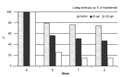
Figure 3.4 En sammenligning af hoved til hale længden af Bombina bombina under metamorfosen. Resultaterne er sammenlignet med længden af de embryoer der lige er blevet overført i et frisk medium (uge 0) og maksimum længden lige før metamopfosen (uge 5). Resultaterne er middelværdier af de to eksperimenter. Middelstandardafvigelsen er mindre end 10%. Mortality During metamorphosis only a slight mortality was found in the control group as shown in figure 3.5. A slight increase in mortality of Bombina bombina previously exposed to 20 μg/l was found. 55.3% of the transferred embryos were still alive just before the beginning of metamorphosis (week 5) whereas this number was reduced to 43.4% after the metamorphosis. The tendency was even more pronounced when Bombina bombina had been exposed to 125 μg/l. In this case 24.5% of the organisms were still alive before the metamorphosis, however, only 14% survived the metamorphosis and as previously described it is assumed that the surviving frogs will only survive for a very short period of time in natural environment.
Figure 3.5 Dødligheden af Bombina bombina under metamorfosen 6,7 and 8 uger efter at embryoer blev overført til pesticid frit medie. Resultaterne er udtryk i % af levende embryoer overført til friskt medie i uge 0. Resultaterne er gennemsnitsværdier af 2 eksperimenter. Standardafvigelsen af gennemsnit er mindre end 10%.
|
