|
The Effect of Esfenvalerate and Prochloraz on Amphibians with special reference to Xenopus laevis and Bombina bombina 3 Results3.1 The effects of vehicles and 6-aminonicotinamide on embryo development of Xenopus laevis and Bombina bombina Egg deposition of both Xenopus laevis and Bombina bombina was about 9-12 h after injection of human chorionic gonadotropin. About 1,000 eggs could be found in the breeding aquarium with Xenopus. However, only about 75-400 eggs were found in the breeding aquarium with Bombina. As for Bombina, the eggs were found on the aquatic vegetation and plastic plants as well as on the small sticks in the aquarium. 3.1 The effects of vehicles and 6-aminonicotinamide on embryo development of Xenopus laevis and Bombina bombina3.1.1 Embryo development in FETAX solutionsControls Only the best fertilised eggs from a given breeding aquarium were used and only normally cleaving embryos were used in these tests. Control mortality could be kept to less than 5% when this procedure was followed. After 96 h, the control group of Xenopus was in the stage 46. A stage 46 larva was recognised by the appearance of the hind limb bud, the coiling of the gut, and the shape of the operculum covering the gills. The best indicator that the larva had attained at stage 46 was the appearance of the hind limb bud. Gut coiling was also easily observed at stage 46 (at stage 45 embryo does not display complete tight gut coiling). At stage 46, the larva was about 9.8 mm in length. After 24 h, the controls of Xenopus were at stage 27, at 48 h (stage 35), 72 h (stage 42), and 96 h (stage 46). Deviations from this standard exposure time were necessary in test with Bombina because the development of the embryos was slower under the same experimental conditions. Thus at 96 h, the embryos of Bombina were at stage 42 corresponding to 72 h in the standard Xenopus test. In embryos of Bombina, the attainment of stage 46 of the controls occurred after 120 h at 24°C. In experiments with Xenopus in which the jelly coat had not been removed, most of the embryos were free of the jelly coat after 24 h at 24°C. Embryos of Bombina, kept at the same temperature, were still in the jelly coat after 24 h and were not free until after about 72 h. Vehicle control The results showed no significant effects of DMSO at a concentration of 1 % v/v, compared with the controls. The mortality and the number of embryos with malformations were less than 5% for both Bombina and Xenopus. The results revealed no reduction of growth rate. At stage 46, the mean length of the embryos of Xenopus and Bombina was 9.8 mm and 9.4 mm, respectively. Vehicle control The results showed no significant effects of acetone at a concentration of 1% v/v, compared with the controls. The mortality and the number of embryos with malformations were less than 5% for both Bombina and Xenopus. The results revealed no reduction of growth rate. At stage 46, the mean length of the embryos was 9.5 and 9.6 for Xenopus and Bombina, respectively. Effect of positive control The results revealed that the effect of 6-aminonicotinamide in the FETAX solutions with Xenopus was comparable with the effect described in the ASTM standards. The EC50 value for malformations after 96 h was 5.3 mg/l and the LC50 value was 2250 mg/l (mean values of 10 experiments; SD of the mean less than 10%). For Bombina, the EC50 value for malformations after 120 h was 4.8 mg/l and the LC50 value was 2450 mg/l (mean values of 10 experiments; SD of the mean less than 10%). For both Xenopus and Bombina, the most conspicuous malformations caused by 6-aminonicotinamide were severe optic cup rupture, displays edema and facial malformations such as ocular edema and foreshortened facial features. In addition, severe heart malformations were also seen. 3.2 Effects of esfenvalerate on Xenopus laevisThe effect of esfenvalerate on malformation, growth inhibition and mortality in Xenopus laevis were tested in the FETAX solution during 96 h of exposure. The effect of esfenvalerate was tested at 1, 2.5, 5, 10, 50, 100 and 150 µg/l and the results were compared with the control group and a DMSO control. The measured concentrations exhibited large variations relative to the nominal values. Since the renewal procedure entailed fresh replacement of the test material every 24 h during the test and the chemical measurement did not reveal a decrease of the pesticide during the 96 h exposure period, the results are expressed as nominal concentrations. The experiments were normally carried out in glass Petri dishes, however, the effect of esfenvalerate was also investigated at concentrations of 1, 10 and 100 µg/l in polystyrene Petri dishes. The results from these experiments were not significantly different from those found in glass Petri dishes. Sumi-Alfa 5 FW The effect of Sumi-Alfa 5 FW was tested at 1, 5, 10 and 100 µg/l (a.i.) to see if the effect was larger when esfenvalerate was added as a formulated product. The results revealed no significant increase in the effect when compared with the experiments where the active ingredient was added with DMSO as a vehicle. 3.2.1 In vivo observationsEsfenvalerate Esfenvalerate affected embryos gradually during the 96 h exposure period and a dose-response relationship was seen. After 24 h, no effect was seen at concentrations up to 5 µg/l, compared with the controls. At 10 µg/l, beginning twisting of the embryos was seen whereas the movements of the controls were more moderate. The intensity of the twisting increased with increasing concentrations of esfenvalerate and at 150 µg/l pronounced twisting was seen in all embryos. After 48 h, no effect was seen at 1 µg/l, however, at 2.5 and 5 µg/l beginning twisting of the embryos was seen. At 10 and 50 µg/l the embryos had constant spasmodic twisting and in 50 µg/l some of the embryos were immobilised. At 100 µg/l, all embryos were immobilised by spasmodic twisting and at 150 µg/l, haemorrhage near the notochord was seen in some of the embryos. After 72 h, beginning twisting was seen in most of the embryos at 1 µg/l and at concentrations above 2.5 µg/l periodic spasmodic twisting was seen. At 10 µg/l, the embryos were immobilised caused by constant spasmodic twisting and at 50, 100 and 150 µg/l many of the embryos had haemorrhage near the notochord and severe eye abnormalities. After 96 h at 1, 2.5 and 5 µg/l, periodic spasmodic twisting was seen in most of the embryos. The intensity increased with increasing concentrations of esfenvalerate. At 10, 50, 100 and 150 µg/l, the embryos were immobilised caused by constant spasmodic twisting and the heartbeat was slow. Many of the embryos had haemorrhage near the notochord, severe eye abnormalities and cardiac edema (the intensity increased with increasing concentrations). 3.2.2 Recordings of malformation, growth inhibition and mortality at the end of the test (96 h)Malformation Most abnormal embryos possessed multiple malformations. However, at 1 and 2.5 µg/l no malformations were seen and the larva were at the normal stage 46 and comparable with the controls in all respects. At a concentration above 2.5 µg/l, the axial malformations increased with increasing concentrations of esfenvalerate. Thus at 5 and 10 µg/l, a relatively mild lateral flexure of the tail was seen. At concentrations above 5 µg/l, optica cup rupture was often seen. Abnormalities of the head and face may be less obvious than other types of malformations, however, at concentrations above 5 µg/l embryos with a sloping forehead and distended mouth were often seen. The head and face regions of these embryos were significantly malformed. The face was extremely flattened and the forebrain was deflected downward along the front of the brow. Heart malformations were seen at concentrations above 5 µg/l. Increasing concentrations of esfenvalerate resulted in increasingly severe heart malformations. Often the heart showed an abnormal expansion of the ventricle just in front of the gut. Gut abnormalities were also seen at concentrations above 5 µg/l where incomplete gut coiling was common. Various degrees of undeveloped gills were also seen at concentrations above 5 µg/l. At higher concentrations (50, 100 and 150 µg/l), severe lateral flexure of the tail was seen. At 50 µg/l, edema was a frequent occurrence. Edema was easily identified and appeared as transparent, swollen, fluid-filled areas. Many embryos exhibited edema in the cardiac region. With increasing concentrations of esfenvalerate embryos displayed increasingly severe abdominal and cardiac edema and at concentrations of 100 and at 150 µg/l optic edema was common.
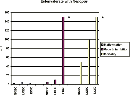
Figure 3-a Effekten af esfenvalerat på Xenopus laevis udtrykt som sublethale defekter, væksthæmning og dødelighed. Testen er udført i overensstemmelse med FETAX standardtesten, og resultaterne udtrykkes ved
angivelse af NOEC, LOEC og EC50/LC50 værdierne efter 96 timers eksponering. Resultaterne er et gennemsnit af 3 forsøg, og SD af middeltallene er mindre end 10%. For yderligere detaljer, jf. Bilag 4. No effects was seen at 2.5 µg/l, however, more than 80% of the embryos had malformations at 5 µg/l and all of the embryos exposed to 10 µg/l and higher concentrations had malformations. The NOEC, LOEC, and EC50 values for malformations were calculated to be 2.0 µg/l, 5 µg/l, and 3 µg/l, respectively (cf. Appendix 4). The most significant malformation observed was cardiac which more than 80% of the embryos had in 5 µg/l. At concentrations above 10 µg/l cardiac, notochord, brain and gut malformations were also common. Growth inhibition After 96 h of exposure to 2.5 µg/l, the mean length of the embryos was measured to 9.7 mm which was comparable with that of the control group. At 5 µg/l and 10 µg/l, the mean length of the embryos was measured to 8.9 and 8.4 mm, respectively. However, no further reduction of the length of the embryos was seen with increasing concentrations of esfenvalerate. Thus, the results revealed that for growth inhibition NOEC was 5 µg/l, LOEC was 10 µg/l and the EC50 was higher than 150 µg/l. (cf. Appendix 4). Mortality No significant mortality was seen at concentrations below 100 µg/l. However, at 100 and 150 µg/l a mortality of about 15% was seen. No significant difference in the mortality rate in the 100 and 150 µg/l samples was observed. The NOEC value for mortality was 50 µg/l and the LOEC value was 100. The LC50 value was higher than 150 µg/l, the highest concentration tested. 3.3 Effects of Prochloraz on Xenopus laevisThe effect of prochloraz on Xenopus was tested at 0.1; 1.0; 1.5; 2.0 ; 3.0, 5.0 and 10 mg/l in FETAX solutions. Acetone was used as a vehicle The measured concentrations exhibited large variations relative to the nominal values. Since the renewal procedure entailed fresh replacement of the test material every 24 h during the test and the chemical measurement did not revealed a decrease of the pesticide during the 96 h exposure period, the results are expressed as nominal concentrations. 3.3.1 In vivo observationsAfter 24 h of exposure, no effect was seen at prochloraz concentrations below 10 mg/l. However, various degrees of edema and blistering were seen at 10 mg/l. After 48 h, some embryos exposed to 1.5 mg/l exhibited mild edema in the cardiac region. With increasing concentrations of prochloraz more and more embryos displayed increasingly severe edema especially optic and cardiac edema. As for embryos exposed to 10 mg/l, the most characteristic malformations were severe optic and cardiac edema. After 72 h, the most characteristic malformation caused by 1.5 mg/l prochloraz was mild edema especially in the cardiac region. In 2 mg/l, embryos with a sloping forehead and edema in especially cardiac and optic region were seen. The heartbeat was slower and decreased when increasing the concentration of prochloraz. At concentrations above 5 mg/l, it seemed like the internal organs of the embryos had more or less loosen from the notochord. Malformations of the gut were also seen and often the gut had coiled into a single loop. Furthermore, severe heart malformations were typical and often the heart was composed of a single beating tube. In 10 mg/l, many embryos (about 20%) were dead and the surviving embryos were immobilised. It was nearly impossible to register the heartbeat in the surviving embryos at 10 mg/l. Edema was a frequent occurrence and may be general (somatic) or regional, e.g., eye (optic), abdominal, cranial or mallar. Many of the embryos also had various degrees of blistering. Blisters were most frequently observed along the dorsal mid-line (fin) area or ventrally near the ventral fin. After 96 h, the above mentioned malformations were seen at concentrations above 3 mg/l. About 60% of the embryos were dead in 5 mg/l and all were dead in 10 mg/l. 3.3.2 Recordings of malformation, growth inhibition and mortality at the end of the test (96 h)Malformation No effect was seen at 0.1 mg/l. However, at 1 and 1.5 mg/l mild rupture in the eye and mild edema in the optic and heart region were seen. Only a few embryos were affected (about 10% increase compared with the controls) in 1.0 mg/l. However, 57% of the embryos had malformations at 1.5 mg/l and all of the embryos exposed to 2 mg/l and higher concentrations had malformations. At 2 mg/l, the above mentioned malformations were normally seen and some embryos had sloping foreheads and their gut were coiled but not to the extent normally expected by stage 46. At concentrations above 3 mg/l most abnormal embryos possessed multiple malformations. The most significant malformations were heart malformations where the heart was often composed of a single straight tube. Often severe abdominal and cardiac edema caused failure to achieve normal gut development. In this case, an almost total failure of the gut to undergo normal coiling was observed. Facial abnormalities generally, though not always, appeared in conjunction with edema of the eyes and head. The most characteristic malformations caused by prochloraz were severe edema, especially optic and cardiac edema. At 5 and 10 mg/l, the surviving embryos normally had edema which may be general (somatic) or regional, e.g., eye (optic), abdominal, cranial, or mallar. Many of the embryos also had various degrees of blistering. Blisters were most frequently observed along the dorsal mid-line (fin) area or ventrally near the ventral fin. In the surviving embryos at concentrations above 5 mg/l it seemed like the internal organs of the embryos were more or less free and loosen from the notochord. Furthermore, severe heart malformations were typical and the heart was often composed of a single tube. Various degrees of incomplete gut coiling were also typical malformations caused by this toxicant. The NOEC value was found to be 0.1 mg/l and the LOEC value was 1.0 mg/l, and the EC50 value was calculated to 1.4 mg/l, cf. Appendix 4.
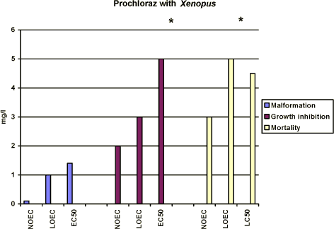
Figure 3-b Effekten af prochloraz på Xenopus laevis udtrykt som sublethale defekter, væksthæmning og dødelighed udført i overensstemmelse med FETAX standard testen. Resultaterne udtrykkes ved angivelse af
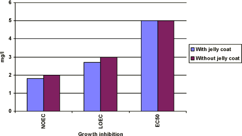
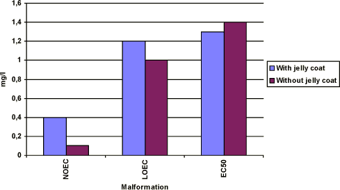
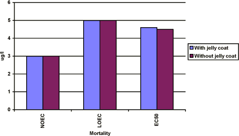
NOEC, LOEC og EC50/LC50 værdierne efter 96 timers eksponering og er et gennemsnit af 3 forsøg. Standardafvigelsen af middeltallene er mindre end 10%. Yderligere detaljer, jf. Bilag 4. Growth inhibition After 96 h of exposure, no effect on the mean length of the embryos was found at concentrations up to 2 mg/l. At 3 mg/l, the mean length of the embryos was measured to 7.7 mm corresponding to about 80% of the controls. At 5 mg/l, the mean length of the embryos was about 76% of the controls. Thus, the results revealed that for growth inhibition NOEC value was 2 mg/l, LOEC value was 3 mg/l and the EC50 value was higher than 5 mg/l. Further details cf. Appendix 4. Mortality No significant mortality was seen at concentrations below 3 mg/l. However at 5 mg/l, a mortality of about 60% was seen. The mortality at this concentration of prochloraz occurred primarily on the last day of exposure. At 10 mg/l, a mortality of 20% was seen after 72 h of exposure and all embryos were dead after 96 h. The NOEC value for mortality was 3 mg/l, the LOEC value was 5 mg/l and the LC50 value was calculated to 4.5 (Further details, cf. Appendix 4). 3.3.3 Comparison of the effects of prochloraz on Xenopus with and without jelly coatIn vivo observations The embryos were free of the jelly coat at all concentrations of prochloraz after 48 h, which is comparable with the control group. No significant difference in either the effects or the time at which the effects were seen at the different concentrations of prochloraz was found when comparing the effect of prochloraz on Xenopus with and without jelly coat. No difference in response in the two groups of embryos was either seen in the control group, acetone control or in the positive control group exposed to 6-nicotinamide. Recordings of malforma-tion, growth inhibition, and mortality at the end of the test (96 h) The results revealed the same effect of prochloraz on growth inhibition, malformation, and mortality, when expressed as NOEC, LOEC and EC50/LC50 values in embryos with and without jelly coat after 96 h of exposure as depicted in figure 3-c to figure 3-e (Further details cf. Appendix 4). Furthermore, the most obvious abnormalities, which were induced by prochloraz in embryos, still retaining their jelly coats, were identical with those seen in embryos whose jelly coats had been removed. Thus, no difference in either the type or seriousness of the malformations was found compared with the effect found in embryos from eggs in which the jelly coat had been removed and then exposed to the same concentrations of prochloraz.
Figure 3-c Effekten af prochloraz på væksthæmningen hos Xenopus laevis. Figuren sammenligner forsøgsresultaterne fra forsøg, hvor man har fjernet gelékappen fra æggene, og hvor man ikke har fjernet gelékappen. Ellers er forsøgene udført i overensstemmelse med FETAX standard testen. Resultaterne udtrykkes ved angivelse af NOEC, LOEC og EC50 værdierne efter 96 timers eksponering og er et gennemsnit af 3 forsøg. Standardafvigelsen af middeltallene er mindre end 10%.
Figure 3-d Effekten af prochloraz på sublethale deformasiteter hos Xenopus laevis. Figuren sammenligner forsøgsresultaterne fra forsøg, hvor man har fjernet gelékappen fra æggene, og hvor man ikke har fjernet gelékappen. Ellers er forsøgene udført i overensstemmelse med FETAX standard testen. Resultaterne udtrykkes ved angivelse af NOEC, LOEC og EC50 værdierne efter 96 timers eksponering og er et gennemsnit af 3 forsøg. Standardafvigelsen af middeltallene er mindre end 10%.
Figure 3-e Effekten af prochloraz på dødeligheden hos Xenopus laevis. Figuren sammenligner forsøgsresultaterne fra forsøg, hvor man har fjernet gelékappen fra æggene, og hvor man ikke har fjernet gelékappen. Ellers er forsøgene udført i overensstemmelse med FETAX standard testen. Resultaterne udtrykkes ved angivelse af NOEC, LOEC og LC50 værdierne efter 96 timers eksponering og er et gennemsnit af 3 forsøg. Standardafvigelsen af middeltallene er mindre end 10%. 3.4 Effects of esfenvalerate on Bombina bombinaThe effect of esfenvalerate on malformation, growth inhibition and mortality in Bombina bombina was tested in the FETAX solution. After 120 h of exposure at 24°C, the embryos in the control group were at stage 46 corresponding to the stage of the controls at the end of the test in the standard FETAX test with Xenopus. The effect of esfenvalerate was tested at 1, 2.5, 5, 10, 50, 100, and 150 µg/l and the results were compared with the control group and a DMSO control. These experiments were made with embryos from eggs where the jelly coat had not been removed. Positive controls experiments with two concentrations of 6-aminonicotinamide were also included in the tests. The results revealed that no effect was seen in the DMSO control group compared with the untreated controls and 56% of the embryos had malformation at 5.5 mg/l 6-aminonicotinamide. A mortality of 43% was found at a concentration of 2500 mg/l. 3.4.1 In vivo observationsEsfenvalerate Esfenvalerate affected embryos gradually during the 120 h exposure in a dose depended manner. After 24 h, no effect of esfenvalerate was seen on the embryos even at the highest concentration compared with the controls. After 48 h, no effect was seen at concentrations up to 5 µg/l, however, at 5 µg/l beginning twisting of the embryos was seen now and then. The spasmodic twisting increased in intensity with increasing concentrations of esfenvalerate and at 150 µg/l the embryos had constant spasmodic twisting. At concentrations from 50 µg/l, the spasmodic twisting of the embryos caused that some of the embryos had already left the jelly coat at this stage. In the control group, only a few embryos had begun to leave the jelly coat after 48 h. At 100 and 150 µg/l, edema was seen in some of the embryos. After 72 h, several of the embryos in the control group began to leave the jelly coat and all the embryos exposed to esfenvalerate concentrations above 50 µg/l were free. Periodic spasmodic twisting was seen in most of the embryos above 5 µg/l and in 50 µg/l constant spasmodic twisting was seen. At concentrations above 100 µg/l, occasionally blistering and cardiac edema appeared. In 150 µg/l, blistering and cardiac edema were frequently seen. The embryos were immobilised by constant spasmodic twisting. After 96 h, all embryos in the control group were free from the jelly coat and beginning twisting was seen at 1 µg/l. The intensity of these spasmodic twisting increased when increasing the concentrations of esfenvalerate. At concentrations above 50 µg/l, the embryos were immobilised caused by constant spasmodic twisting and occasionally blistering and cardiac edema appeared. At concentrations above 100 g/l, blistering and edema were frequently seen as well as head and brain malformations. The heartbeat was very slow and severe heart malformations were normally seen. After 120 h, spasmodic twisting was seen at 1 µg/l. At concentrations above 50 µg/l, the embryos were immobilised caused by constant spasmodic twisting and blistering and edema were often seen. At concentrations above 100 µg/l, blistering and edema were frequently seen as well as head and brain malformations. The heartbeat was very slow and severe heart malformations were seen at 100 and 150 µg/l. 3.4.2 Recordings of malformation, growth inhibition and mortality at the end of the test (120 h)Malformation At concentrations up to 2.5 µg/l, no malformations were seen and the larva were at the normal stage 46 and comparable with the controls in all aspects. Above a concentration of 5 µg/l, gut abnormalities were often seen indicated by a loose gut coiling. At 10 µg/l in addition to gut abnormalities, a relatively mild flexure of the tail was normally seen. Embryos with a sloping forehead and distended mouth were often seen. The head and face region of these embryos were malformed. The face was flattened and the forebrain was deflected downward along the front of the brow. Heart malformations were also observed at concentrations above 10 µg/l. Increasing concentrations of esfenvalerate resulted in increasingly severe heart malformations. Often the heart showed an abnormal expansion of the ventricle just in front of the gut. At 50 µg/l, blistering and edema were common and various degrees of undeveloped gills were seen, too. About 10% of the embryos had malformations at 10 µg/l and all of the embryos exposed to 100 µg/l and higher concentrations had malformations. The EC50 value for malformations was calculated to be 29 µg/l (Further details cf. Appendix 4). The most significant malformation observed was flexure of the tail and cardiac.
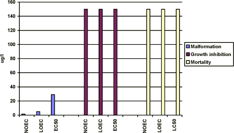
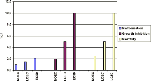
Figure 3-f Effekten af esfenvalerat på Bombina bombina udtrykt som sublethale defekter, væksthæmning og dødelighed udført i overensstemmelse med FETAX standardtesten. Resultaterne udtrykkes ved angivelse af NOEC, LOEC og EC50/LC50 værdierne efter 120 timers eksponering og er et gennemsnit af 3 forsøg. For yderligere detaljer, se bilag 4. Growth inhibition After 120 h of exposure to concentrations up to 150 µg/l, the mean length of the embryos was measured to about 9.4 mm which was comparable with the length of the control group. Thus, the results revealed that for growth inhibition NOEC, LOEC and the EC50 valued were higher than 150 µg/l. Mortality No significant mortality was seen at concentrations up to 150 µg/l. The NOEC, LOEC, and LC50 values were all higher than 150 µg/l. 3.5 Effects of prochloraz on Bombina bombinaThe effect of prochloraz on Bombina was tested at 1.0, 1.5, 2.0, 5.0 and 10 mg/l in FETAX solution at 24°C. Acetone was used as a vehicle and an acetone control was therefore included in the test. The results revealed no significant effect in either growth inhibition, malformation or mortality when the acetone control was compared with the control group. Addition of 5.5 mg/l 6-aminonicotin-amide induced 57% malformation of the embryos and addition of 2500 mg/l induced 42% mortality after 120 h of exposure. 3.5.1 In vivo observationsAfter 24 h of exposure, no effect was seen at any prochloraz concentrations. After 48 h, no effect was seen at prochloraz concentrations up to 2 mg/l. However, embryos exposed to 5 and 10 mg/l showed signs of mild edema, especially in the optic and cardiac region. After 72 h, the embryos began to leave the jelly coat both in the control group and in samples exposed to prochloraz. However, at 10 mg/l prochloraz only a few embryos were free. No effect was seen at concentrations up to 2 mg/l. The most characteristic malformations caused by 2 mg/l prochloraz were mild edema, especially in the cardiac region. In 5 and 10 mg/l, embryos with a sloping forehead and edema especially in cardiac and optic region were seen. After 96 h, all embryos were free. Embryos exposed to 1.5 and 2 mg/l showed signs of edema especially in the cardiac region. In 5 and 10 mg/l, embryos with a sloping forehead and edema in especially cardiac and optic region were seen. Severe heart malformations were typical and the heart was often composed of a single beating tube. The heartbeat was slower at these concentrations and at 10 mg/l it was nearly impossible to observe heartbeat. The surviving embryos in 5 and 10 mg/l were immobilised (did not react on mechanical stimuli). Edema was a frequent occurrence and may be general (somatic) or regional, e.g., eye (optic), abdominal, cranial, or mallar. Many of the embryos had also various degrees of blistering. Blisters were most frequently observed along the dorsal middling (fin) area or ventrally near the ventral fin. Malformations of the gut were also seen. After 120 h, no effect was seen at 1 mg/l but at 1.5 mg/l edema was seen and at 2 mg/l the embryos were immobilised and edema was a frequent occurrence especially in eye (optic), abdominal, cranial, and heart. In 5 and 10 mg/l, the above (after 96 h) mentioned malformations were seen in a more pronounced manner. About 10% of the embryos were dead in 5 mg/l and 25% at 10 mg/l. 3.5.2 Recordings of malformation, growth inhibition and mortality at the end of the test (120 h)Malformation No effect was seen at 1 mg/l. However, at 1.5 mg/l mild edema in the heart region was seen. Only a slight effect (20% increase compared with the controls) was found at 1.5 mg/l, however, about 50% of the embryos had malformations at 2 mg/l and all of the embryos exposed to 5 and 10 mg/l had malformations. At 2 mg/l, the edema especially in the heart region was often seen and some embryos had a sloping forehead. At 5 and 10 mg/l, most embryos possessed multiple malformations, where the most significant was heart malformations. Often severe abdominal and cardiac edema caused failure to achieve normal gut development. Facial abnormalities generally, though not always, appeared in conjunction with edema of the eyes. The most characteristic malformations caused by prochloraz were severe edema especially optic and cardiac edema. Many of the embryos also had various degrees of blistering. Blisters were most frequently observed along the dorsal middling (fin) area. It seemed that the internal organs of the embryos were more or less free and loosen from the notochord. The NOEC value was found to be 1 mg/l and the LOEC value was 1.5 mg/l. The EC50 value for malformations was calculated to be 2.1 mg/l (Further details cf. Appendix 4). The most significant malformations observed were cardiac, brain, edema, and gut malformations.
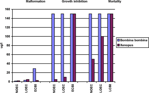
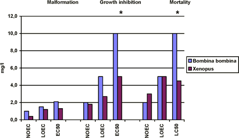
Figure 3-g Effekten af prochloraz på Bombina bombina udtrykt som sublethale defekter, væksthæmning og dødelighed. Resultaterne udtrykkes ved angivelse af NOEC, LOEC og EC50/LC50 værdierne efter 120 timers eksponering og er et gennemsnit af 3 forsøg. Standard afvigelsen af middeltallene er mindre end 10%. For yderligere information se Bilag 4. Growth inhibition After 120 h of exposure, no effect on the mean length of the embryos was found at concentrations up to 2 mg/l. At 5 mg/l, the mean length of the embryos was measured to 7.4 mm corresponding to about 80% of the controls. At 10 mg/l, the mean length of the embryos was about 70% of the controls. Thus, the results revealed that for growth inhibition NOEC was 2 mg/l, LOEC was 5 mg/l, and the EC50 was higher than 10 mg/l. Mortality No significant mortality was seen at concentrations below 5 mg/l. However, at 5 mg/l a mortality of about 10 % was seen. The mortality at this concentration of prochloraz occurred primarily on the last day of exposure. At 10 mg/l, a mortality of 25% was seen after 120 h of exposure. The NOEC value for mortality was 2.5 mg/l, the LOEC value was 5 mg/l and the LC50 value was more than 10 mg/l. 3.6 Comparison of the effects of esfenvalerate and prochloraz on Xenopus and Bombina3.6.1 Comparison of the effects of esfenvalerate on Xenopus and BombinaWhen a comparison of the effects of esfenvalerate on malformation in Xenopus and Bombina was made the results revealed that the NOEC and LOEC value in both species were 2.5 µg/l and 5 µg/l, respectively. However, a significant difference between the two species was found at 5 µg/l. In tests with Xenopus, more than 80% of the embryos had malformations at 5 µg/l and all of the embryos exposed to 10 µg/l had malformations while only about 10% of the embryos exposed to 10 µg/l had malformation in tests with Bombina. However, all embryos exposed to 100 µg/l had malformations. The EC50 value was calculated to be 3 µg/l and 29 µg/l for Xenopus and Bombina, respectively. However, an effect of esfenvalerate was seen at a concentration of 1 µg/l in the in vivo observations in both Xenopus and Bombina. The most characteristic effect of esfenvalerate on living embryos of both species was the spasmodic twisting which at the higher concentrations caused a complete immobilisation of the embryos. The most significant malformations observed after the end of the tests and fixation of the embryos were also identical in the two species and included cardiac, severe lateral flexure, edema, notochord, brain and gut malformations. The effect of esfenvalerate on growth seems to be different in the two species. The results revealed that in Xenopus addition of 10 g esfenvalerate only caused a reduction of about 15% of the embryos' mean length compared with the controls. No further reduction of the length of the embryos was seen when increasing the concentrations of esfenvalerate up to 150 µg/l. Thus, the results revealed that for growth inhibition NOEC was 5 µg/l, LOEC was 10 µg/l and the EC50 value was higher than 150 µg/l. In experiments with Bombina, esfenvalerate did not affect the mean length of the embryos at any concentrations up to 150 µg/l. Thus, the NOEC, LOEC and EC50 values were all higher than 150 µg/l. In Xenopus, a significant mortality of about 15% was seen both at 100 and 150 µg/l compared with the control group, but the LC50 value was higher than 150 µg/l. For Bombina, the results revealed that no significant mortality was seen at concentrations up to 150 µg/l compared with the controls.
Figure 3-h En sammenligning af effekterne af esfenvalerat på Xenopus og Bombina udtrykt ved sublethale defekter, væksthæmning og dødelighed. Resultaterne udtrykkes ved angivelse af NOEC, LOEC og EC50/LC50 værdierne efter 96 og 120 timers eksponering for henholdsvis Xenopus og Bombina. Efter denne eksponeringstid er embryonerne i de respektive kontrolgrupper nået til stadie 46. Resultaterne er et gennemsnit af 3 forsøg. Standardafvigelsen af middeltallene er mindre end 10%. 3.6.2 Comparison of the effects of prochloraz on Xenopus and BombinaWhen a comparison of the effect of prochloraz on malformation in Xenopus and Bombina was made, the results revealed that no effect was found at concentrations below 1 mg/l. However at 1 mg/l, about 10% of the embryos of Xenopus had malformations while no effect was seen in Bombina. A significant difference between the two species was found at 1.5 and 2 mg/l. Thus in tests with Xenopus, about 60% of the embryos had malformations at 1.5 mg/l and all of the embryos exposed to 2 mg/l and higher had malformations while only about 20% of the embryos exposed to 1.5 mg/l had malformation in tests with Bombina and about 50% of the embryos had malformations at 2 mg/l. All embryos exposed to 5 mg/l and higher concentrations had malformations. The EC50 value was calculated to be 1.3 and 2.9 mg/l for Xenopus and Bombina, respectively. The most characteristic effect of prochloraz on living embryos of both species was severe heart malformations. At the higher concentrations, the heartbeat was nearly impossible to observe and the surviving embryos were so damaged that they did not react on mechanical stimuli. After the end of the test, the most significant malformations observed were also identical in both species and included cardiac, brain, edema, and gut malformations. The effect of prochloraz on growth in both species seemed to be comparable and no effect was seen at concentrations below 2 mg/l. However, in Xenopus addition of 3 mg/l prochloraz caused a reduction of the mean length of the embryos of about 20% as compared with the controls and at 5 mg/l the mean length was reduced with about 25%. In experiments with Bombina 5 mg/l prochloraz caused a reduction of about 20% as found in experiments with Xenopus at 3 mg/l. About 75% of the embryos were still alive after 120 h exposure to 10 mg/l prochloraz and a reduction of the mean length of the surviving embryos was about 30% compared with the controls. In Xenopus, no significant mortality was seen at 3 mg/l. However, about 56% of the embryos were dead at 5 mg/l and all embryos were dead after 96 h exposure to 10 mg/l. For Bombina the results revealed that no significant mortality was seen at concentrations up to 2 mg/l. At 5 mg/l, a mortality of about 10% was seen and at 10 mg/l about 25% of the embryos were dead at the end of the experiments.
Figure 3-i En sammenligning af effekterne af prochloraz på Xenopus og Bombina udtrykt ved sublethale defekter, væksthæmning og dødelighed. Resultaterne udtrykkes ved angivelse af NOEC, LOEC og EC50/LC50 værdierne efter 96 og 120 timers eksponering for henholdsvis Xenopus og Bombina. Efter denne eksponeringstid er embryonerne i de respektive kontrolgrupper nået til stadie 46. Resultaterne er et gennemsnit af 3 forsøg. Standardafvigelsen af middeltallene er mindre end 10%. 3.6.3 The effects of esfenvalerate and prochloraz on TI on Bombina and XenopusThe TI is defined as the 96 h LC50 divided by the 96 h EC50 (malformation) and is a measurement of developmental hazard. Table 3-a Teratogen index (TI) for de to pesticider esfenvalerate og prochloraz.
The results from table 3-a revealed that in this test esfenvalerate as well as prochloraz can be considered as a teratogen according to this test.
|
