Dermal absorption of pesticides - evaluation of variability and prevention
2 Human skin structure and function
The skin is a complicated human organ (Figure 1), which is continuously exposed to chemicals, mechanical injury, micro-organisms, UV light, temperature fluctuations and water loss. The most important function of this organ is to minimize unintended water loss and maintain homeostasis and to act as a barrier against these exposures. As the largest single organ of the body, the skin accounts for more than 6% of the body mass; on average about 5 kg covering 1.8m² depending on height and weight (Roberts MS & Walters KA, 1998b;Thestrup-Pedersen K et al., 1993). The cutaneous blood circulation comprises 5-10% of the total cardiac output (Johnson et al., 1986).
The skin is a heterogeneous organ, holding a number of layers as well as appendages – hair follicles, sebaceous glands and sweat glands. Different body regions show different skin thickness and composition of the stratum corneum. The skin can be divided into an outer region - the upper epidermis and an inner region - the lower more vascular dermis. The viable epidermis can metabolize chemicals that infiltrate the stratum corneum. The dermis offers physiological support for the avascular epidermis and is the source of nutrition.
2.1 Epidermis
The epidermis can be divided into four layers (from inner to outer layer):
2.1.1 Stratum basale (germinativum)
Following the life circle of the keratinocytes they start out as a single layer of columnar basal cells attached to the basement membrane by hemidesmosomes creating the stratum basale (Roberts MS & Walters KA, 1998b). They consist of metabolically active cells, which continuously proliferate and undergo mitosis, causing the older cells to move out towards the surface.
2.1.2 Stratum spinosum
Stratum spinosum lies on top of the stratum basale and is also known as the prickle layer. These cells adjacent to the basal layer generate lamellar granules that soon after fuse with the cell membrane to release the neutral lipids thought to generate a barrier to penetration through the epidermis. This part of the epidermis consists of many layers of irregular cells connected to the surrounding cells by desmosomes. As the cell layers move outwards they flatten, and the granules reflect the border between this stratum and the overlying stratum granulosum.
2.1.3 Stratum granulosum
The cells in stratum granulosum are even more flattened than in the previous layer. They contain an increasing amount of keratin as further keratin differentiation occurs in this layer. The most characteristic elements are the intracellular granules. They hold many different components that all together play an important role in the keratinisation process of the cells. Enzymes degrade the viable cell components such as nuclei and intracytoplasmatic organelles. The cells in the stratum granulosum also contain large amounts of filaggrin, a protein thought to serve in bundling keratin. Filaggrin is an element in the keratinisation process which together with lipids helps to create a protective barrier against penetrating substances. Mutations in the filaggrin gene have proved to create an impaired barrier function as it gives a varying degree of abnormal skin conditions (Palmer et al., 2006). This will be described in the section dealing with the barrier function.
2.1.4 Stratum corneum
Stratum corneum, also known as the horny layer, develops from immature, columnar, keratinocytes that, as they move from the basal layer and by that the source of nutrition to the stratum corneum, become flat, keratin-filled and dead cells. On the way to the surface, they loose the nucleus and the capacity for metabolic activity. From the body surface the dead cells are constantly shedded and therefore the skin barrier is continuously renewed. The total cell turnover of epidermal cells is between 17 and 71 days depending on the anatomical site (Maibach H & Patrick E, 2001). The stratum corneum provides almost all mechanical strength to the epidermis.
The stratum corneum varies in thickness (10-600mm) depending on the area of the body and the physical interaction that the skin is exposed to in daily life. Plantar and palmar callus can be 400-600mm thick compared to 10-20 mm for the back, arms, legs and abdomen (Scheuplein & Blank, 1971).
The stratum corneum consists of several flattened, hexagonal and cornified cells stacked on top of each other, usually 15–20 cells thick. It is a heterogeneous structure containing about 40% protein (mainly keratin, a disulfide cross-linked linear polypeptide), 40% water (depending on humidity) and about 15 to 20% lipids (principally triglycerides, free fatty acids, cholesterol, and phospholipids) (Michales AS et al., 1975). The lipid is concentrated in the extracellular phase and the protein in the intra- and extracellular phase. Stratum corneum’s protein-rich corneocytes embedded in a matrix of ceramides, cholesterol, and fatty acids, and smaller amounts of cholesterol sulphate, glucosylceramides and phospholipids were described by Elias (1983) and Bouwstra (2006) as the “Brick and mortar” model (Elias, 1983;Bouwstra & Ponec, 2006) and later by Forslind (1997) in a more detailed model “the domain mosaic model” (Forslind et al., 1997). Because of the contents of lipids it has a low permeability to many agents and a protective function for the individual. The bricks (keratinocytes) act as a hydrophilic membrane and are almost impenetrable to water and by that a regulating mechanism to limit transepidermal water loss. The mortar (lipids) acts as a regulating mechanism for hydrophobic penetration.
A previous study has indicated that a too high as well as a too low lipophilicity limits the skin penetration of a substance (Nielsen JB, 2004), but further research needs to be done to determine an interval in relation to lipophilicity where the most efficient absorption could be expected. This information will potentially allow an improved prediction of the ability of new chemicals to penetrate the skin.
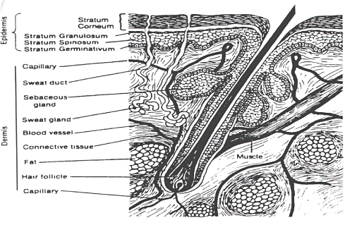
Figure 1: Skin structure.
2.2 Dermis
The epidermis rests on the much thicker dermis (2000mm), which again lies above the subcutaneous fat. Dermis consists of collagenous fibres (70%), providing a scaffold of support and cushioning, and elastic connective tissue, providing elasticity, in a semigel matrix of mucopolysaccharides. The dermis also embeds mastcells (part of the immune system), melanocytes (produce pigment) and the fibroblasts (produce the connective tissue). Moreover, the dermis also carries the blood, nerve and lymph supply. This vascular network of microcirculation supports the skin with nutritients and acts as a heat regulator besides absorbing penetrated chemicals and transporting them to the systemic circulation (Guy et al., 1987). The nerves respond to pressure and temperature. The lymphatic system is an important component of the skin in regulating its interstitial pressure, mobilization of defence mechanism, and in waste removal (Roberts MS & Walters KA, 1998a). While blood flow establishes the clearance of small solutes such as water and other small particles, lymphatic flow is an important determinant in the dermal removal of larger solutes (Cross & Roberts, 1993).
2.3 Skin appendages
The skin contains different appendages such as hair follicles with its sebaceous glands, eccrine glands, apocrine glands and nails. Each has different functions that provide the skin with its many abilities to protect the human body.
The hair is a keratinised structure that acts as protection and is distributed on most of the body. The hair cycle is controlled by temperature, light, nutrition and hormones. The hair follicles are rooted in the dermis, sometimes extending into the hypodermis.
The sebaceous glands have a protective function. They secrete sebum, which acts as lubricant to waterproof the skin and prevent it from drying. Androgenic hormones stimulate the sebaceous glands.
The eccrine glands are sweat glands that are found all over the body with a majority on the soles, palms and axillae. The sympatic nerves activate them and the function is to regulate body temperature.
The apocrine glands are found in the axillae, the nipples and the anogenital area and secrete proteins, lipids and lipoproteins. They are also sweat glands, but with a different function and morphology than the eccrine glands. Both types of glands are activated by the sympathetic nerve system (Roberts MS & Walters KA, 1998b).
Version 1.0 May 2009, © Danish Environmental Protection Agency