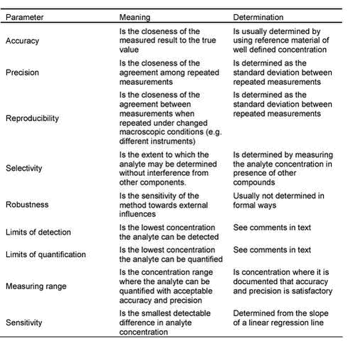
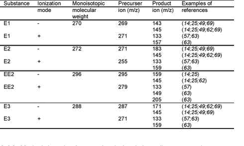
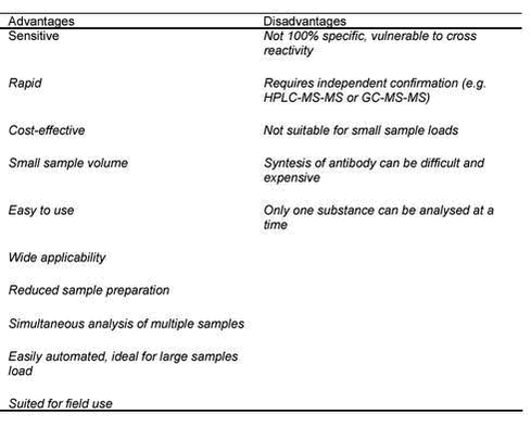
Evaluation of Analytical Chemical Methods for Detection of Estrogens in the Environment3 Analytical methodologies3.1 Conceptual description of analytical methods3.2 Validation of methods for analysis of estrogens 3.3 Sample handling 3.3.1 Sample collection, preservation and handling 3.3.2 Sample preparation 3.4 Analytical detection methods 3.4.1 Methods based on gas chromatography (GC-MS and GC-MS-MS) 3.4.2 Methods based on high performance liquid chromatography (HPLC) 3.4.3 Methods based on Immunochemical techniques (immunoassay) 3.4.4 Other techniques This chapter describes the different techniques used for analysis of estrogens. The methods are described from a technical and chemical perspective. Giving an overview of the publications describing the methods is also an objective of this section. 3.1 Conceptual description of analytical methodsIt is important to realise that an analytical chemical method consists of a number of individual steps that can be divided in four groups as shown as in Table 3.1. This table also give examples of problems that should be considered under each step. An optimal chemical analysis is only achieved if the behaviour of the substances to be analysed (the analytes) is well understood in each of these steps. It is important to realise that an error made in one of these steps (e.g., during sampling) may have consequences for the whole analytical method. Table 3.1: 3.2 Validation of methods for analysis of estrogensThe ability to provide timely, accurate, and reliable data is central to any method for analysis of chemicals. Therefore method validation should be an integrated part of the process of developing analytical methods. A number of authorities and organisations have published detailed guidelines stating how method validation should be performed (see e.g., (40)), for an overview). In the current context it is important to note that these descriptions cover many fields of analytical chemistry and contain many details. However, all the various guidelines state that it is important to define the intended purpose of an analytical method and that method validation only needs to prove that the method is acceptable for this purpose. In environmental analysis this is particularly important, as the need of high precision may be limited. For steroid estrogens specifically, the purpose of analytical methods often can be limited to reliably detecting whether the substances are present above a certain concentration level or not. Thus in this particular case, only limited method validation is needed. A number of validation parameters are defined in order to describe the quality of the entire analytical process. These parameters are determined on the basis of a validation procedure, including the analysis of a range of samples in matrix. It is important to stress that the validation should cover all the steps of the analytical procedure (including sampling, storage and preparation). An overview of the validation parameters is given in Table 3.2. Table 3.2: The accuracy, precision, and reproducibility are all expressing the confidence one may attach to an analytical result. For several reasons, these parameters are of considerable importance in the analysis of estrogens in the environment. Particularly, when samples of different origin are analysed. Therefore, it is important to stress that these parameters, such as the standard deviation, confidence intervals or similar quantifiers, should be specified in the reporting and assessment of any analytical result. As estrogens belong to the group of steroids, which are all very similar chemical compounds, the selectivity problems encountered are often due to interference from other steroids in the matrix. In the current context, the selectivity problems are considered severe making it necessary to document the selectivity of analytical methods. Due to limited selectivity, several authors who published monitoring results are suspected of over-estimating the concentrations of steroid estrogens in the environment. An important example is the work by Kolpin and co-workers (41) who reported concentrations that were higher than worst-case estimates could substantiate (42;43). The selection of methods for analysis of steroid estrogens in environmental samples is most often dictated by the limits of detection and quantification since extreme sensitivity is required in most cases. The determination of the limits of quantification and detection (LOQ and LOD) can be made in several ways and has therefore been subject for discussion (44). All approaches however, depend on an estimate of precision at or near zero concentration. In practice LOD and LOQ are expressed as the concentration where the relative standard deviations of replicate samples is below a certain level (typically within 95% confidence limits). Alternatively LOD/LOQ is defined on basis of a comparison of the strength of the analytical signal with the strength of the background signal (signal to noise ratio). 3.3 Sample handling3.3.1 Sample collection, preservation and handlingVery few articles in the literature address sample collection, preservation and handling of natural estrogens in environmental matrices. Existing records reveal that both discrete and composite samples of waste water influents, effluents and occasionally, partially treated STP water are collected, analysed and reported. Sampling periods ranging from 6 hours to 5 days have been used to collect composite samples. In many instances discrete samples have been colleted. Grab (discrete) samples do not, in general, seem appropriate for assessing the presence of estrogens and xeno-estrogens in influents and effluents, particularly if the aim of the study is to evaluate the performance of the STP plant. Despite this many of the studies in the literature were performed with grab samples of effluents alone, and no samples of influents were collected at all. Some papers have justified the use of discrete samples in preference to bulked composite samples by remarking that the stability of estrogen compounds in waste water was, at that time, unknown. One study by Baronti et al. (25) has investigated the stability of estrogens in treated and untreated water samples. Baronti and co-workers showed in their study that the best storage strategy passed the field samples through the extraction cartridge (C18 or Carbograph-4), washed the cartridge with a methanolic water solution (10%), and stored the cartridge at -18°C. Under these conditions, which provide a practical way to store many samples in extensive monitoring programs, no significant loss of estrogens was observed after storage for 60 days. An alternative but less secure procedure is to store the water in bottles preserved with 1 % aldehyde at 4°C. In the same paper Baronti and co-workers reported that there were no significant losses of the estrogens after 28 days when the samples were preserved, but severe losses occurred when the samples were not preserved. Other studies have used methanol, sulphuric acid or mercuric chloride to preserve the samples, however, methanol should under no circumstances be selected as the preservation chemical as this may influence the deconjugation af steroids in the sample. Methanol will always increase the degradation of the estrogens in the matrix in question. Data indicative of the potential degradation of estrogens in unpreserved water for storage periods shorter than 7 days, for instance 24-48 h, were, unfortunately, not available. Freezing of unpreserved samples at -20 °C has been used in a few studies. Unfortunatly these studies have not investigated the loss of estrogenzzs during storage. From a preliminary study of the estrogen concentration in samples of effluents from 20 Danish STPs, our experience indicates that the estrogenconcentration decreased significantly in unpreserved water samples within 48 hours (45). The volume of sample processed depended mainly on the sensitivity of the technique used for the final analysis and varied from 50 mL extracted and analysed by radioimmunoassay by Shore et al (46) to 20 L (8) or even 80 L, extracted by liquid-liquid partition and analysed by gas-liquid chromatography by Tabak et al (47). It is not advisable to extract more than approximately 5 L with existing sample preparation methods because greater volumes only create other problems, e.g., extracts with a high load of humic acid. The literature (e.g., Ternes et al. (26) provides only a few examples of how to sample soil, manure or sludge (solid phase of sludge). Care should be taken to investigate the loss of analyte in these matrices by performing recovery studies on the effects of time, handling methods and sample preservation techniques. 3.3.2 Sample preparationSection 3.3.2 primarily discusses sample preparation methods presented in the papers referred to in appendices 1 to 4. 3.3.2.1 Sample prep and cleanup for chemical analyses and bioassays Filtration Extraction SPE cartridges Sample-loading flow rates varied greatly among applications but were usually between 0.5 and 70 mL/min. Subsequent drying of the cartridge with either nitrogen or air is a common practise with no reported analyte loss. Purification Evaporation 3.3.2.2 Deconjugation techniques using enzymatic hydrolysis In many cases non-conjugated as well as conjugated estrogens are present in environmental samples. For several reasons detection and measurement of these constituents may be unnecessary. In such cases, deconjugation of all the estrogens in the sample has been suggested. Estrogen conjugates in waste water can be quantified by including an hydrolysis step which converts the conjugated forms into the active hormones during the sample preparation process. This step is necessary if final analysis is performed with GC-MS or GC-MS-MS. Both LC-MS and Immunoassay techniques can be used for direct analysis of conjugates although the methods have the same drawbacks as for the unconjugated compounds. Comparing the results of thes two methods allows one to simultaneously determine the concentration of both free and conjugated forms. The LC-MS method makes a direct measurement of conjugates and avoids the hydrolysis step. This is a clear advantage. 3.3.2.3 The use of standard and internal standards In order to obtain accurate values for the estrogen concentrations, the standard used should be as pure as possible (near 100%). In any case, the amount of estrogen present in a certain quantity of the standard material should be known exactly. Therefore, certified reference material should be used if available. 14C-label has often been employed (located in the steroid skeleton) as an internal standard, which has the advantage that no label exchange occurs, but a major disadvantage is that its radioactivity makes it a health hazard risk. Another disadvantage of 14C- labelled steroids is the general inclusion of appreciable amounts of unlabelled material. Therefore at present most researchers employ isotopically labelled steroids in which some (at least two) hydrogen atoms are replaced by deuterium atoms at free exchange positions or alternatively, at least two 12C atoms are replaced by 13C. Besides being radioactively stable these internal standards contain only a few percent unlabelled compounds. Precise measurement of the amount of the unlabelled and labelled steroids is of crucial importance, as this eventually determines the accuracy and precision of the end result. The analytical balance and pipettes used should be carefully calibrated and their tolerance known. In all relevant papers much attention has been paid to this subject. In the hands of experienced personnel this stage in the analytical procedure can be carried out in such a way that it contributes no more than 0.1-0.2 % of the total error in the final result. The internal standard should be added as the first analytical step, mostly as an alcoholic solution; it is important that good equilibrium is reached before extraction and that the amount of alcoholic solution added does not result in precipitation of other constituents. 3.3.2.4 Comparison of sample prep methods use for bioassays and for chemical analysis Sample preparation for bioassays and chemical analysis is in principal identical. The more clean-up of the sample the less probability that many false-positive results will occur. High loads of suspended solids will result in undesired adsorption on to antibodies. The highest level of sample prep and cleanup is especially important when using bioassays. For some of the most sophisticated GC-MS-MS methods less clean-up may be acceptable. 3.4 Analytical detection methodsEstrogens are detected using several different detection methods. The purpose of the current section is to describe methods published in the scientific literature. It is important to stress that the techniques used for detection of estrogens are used in other contexts than environmental analysis and therefore a vast amount of literature related to other fields (see e.g., (52)) exists, but these will not be covered here. Currently, most of the work related to the analysis of steroid estrogens in the environment has been made on surface water or sewage influent or effluent. Therefore, only for these matrices have analytical methods with satisfactory method validation for detection of steroid estrogens been documented. This will be reflected in the current section that primarily deals with analytical methods for such matrices. 3.4.1 Methods based on gas chromatography (GC-MS and GC-MS-MS)3.4.1.1 General information In gas chromatography (GC) the compounds to be analysed are vaporised and eluted by a stream of gas as a mobile phase through the column. The mobile phase is used only as a carrier gas so that interactions of the mobile phase with the analyte are of no significance. The analyte is normally dissolved in a liquid and GC is normally used primarily for volatile organic compounds. The predominant separation principle is then the partition of substances between the liquid stationary phase and the gaseous mobile phase. GC-MS is the most popular of all hyphenated techniques for gas chromatography, and the combination of a powerful separation technique with the high degree of structural information provided by the mass spectrometry (MS) has made GC-MS the workhorse of trace analytical laboratories. This combination gives the possibility of combining an automated separation on the GC with structural information (masses) on the MS. The most popular mass analyser is the quadrupole mass filters that allow high scanning speeds up to a transmission range of m/z equal 2000. GC-MS-MS is the hyphenated technique combining the GC with a tandem mass spectrometry (MS-MS) (triple quadrupole). MS-MS is any general method involving at least two stages of mass analysis either in conjugation with a dissociation process or a chemical reaction that causes a change in the mass or charge of an ion (see below). In the most common MS-MS a first analyser is used to isolate a precursor ion, which then undergoes a fragmentation, either spontaneously or by some activation, to yield product ions and neutral fragments. A second spectrometer analyses the product ions. By using a MS-MS instrument the selectivity of the analysis is increased as not only is a specific mass used for quantification, but this specific mass can be related to a specific fragmentation of product ions. If the analyte is part of a complicated matrix this will reduce the matrix interference. If single GC-MS is used for analysing compounds in complicated matrices, such as treated or un-treated waste water, rules for identification must be set up. These rules might include matching of retention time, presence of molecular ion of target compound, presence of at least two additional qualifier ions and matching of ion ratios within 50% for the two qualifier ions. A special version of the MS-MS instruments is the ion-trap MS-MS. An ion trap can be imagined as a quadrupole bent on itself in order to form a closed loop. This allows the instrument to trap a large number of molecules for ionization, thereby increasing the sensitivity of the instrument. These instruments have regularly been applied in this field. For more information on MS detectors see the chapter on liquid chromatography based techniques. 3.4.1.2 Ionisation methods Although various ionization methods are available, electron impact (EI) and chemical ionization (CI) are the most common for general use in GC-MS analysis. Of these two techniques EI is by far the most widely used. EI ionization produces fragment rich mass spectra that may provide structural information. In EI sample molecules entering the ion source from the gas chromatographic column, are ionized by thermal electrons emitted from a tungsten or rhenium filament (the cathode) and accelerated towards the anode. The electrons collide with the sample molecules, transferring part of the kinetic energy of the electrons to the molecules. This causes excitation, fragmentation, and ionisation. As the distribution of the internal energy directly affects the appearance of the mass spectra, and is strongly dependent on the electron beam energy, (Eel), it is usually fixed at a standard value of 70 eV. Spectra are compiled in libraries and used for identification of compounds via a search procedure. 3.4.1.3 The use of GC for analysing natural estrogens The analytical determination of natural estrogens in environmental matrices has been dominated by the use of GC-MS and GC-MS-MS. Both conventional MS and MS-MS (ion trap and triple quad.) detection are accomplished in the EI mode of ionization. The use of NCI and PCI has also been reported, however, it is important to note that only GC combined with MS and tandem MS, respectively, provide sufficient selectivity and inherent sensitivity to analyse for natural estrogens in complicated matrices such as treated waste water, sludge, manure or soil. The major difference between single MS and MS-MS is in the selectivity of the analysis (see example in section 5). Interference from the matrix may be a major problem with single MS. This problem is especially acute for EE2, where measured concentrations are sometimes higher than anticipated suggesting that this difference may be due to interference by natural organic matter. Using MS-MS this interference may be reduced by using one or more daughter ions for quantification. In most studies the instrument has been operated at 70 eV, in the selected ion monitoring (SIM) mode. The analysis is conducted after sample derivatization (see below). The use of derivatization agents in sample preparation for GC analysis is one of the major drawbacks of using GC for analysing natural steroids (see below). An overview of recent publications presenting GC detection based methods is given in Appendix 3. 3.4.1.4 GC-methods with non-MS detectors The use of GC for analysis of natural estrogens in complicated environmental matrices without the hyphenation of an MS instrument is not recommended and therefore not treated further in this work. 3.4.1.5 Detection limits The detection limits achieved with the different methods employing GC-MS or GC-MS-MS as final analytical techniques were in the range of 0.5 – 74 ng/L and 0.1 – 24 ng/L, respectively. 3.4.1.6 Capillary Columns GC separation is performed with a variety of capillary columns using helium as carrier gas, with temperature programs from approximately 45 to 300 °C. Sample volumes of 1 to 4 µL extracts are injected in the splitless mode. 3.4.1.7 Derivatization agents In order to improve the stability of the compounds and the sensitivity and precision of the GC-MS or GC-MS-MS analysis derivatization agents are always used. Several derivatization agents such as bis-(trimethylsilyl)-triflouroacetamide, N-methyl-N-(tert.)-butyl-dimethylsilyl-triflouroacetamide (MTBSTFA) and heptaflouro-butyric anhydride, have been used depending on the choice of ionisation technique. The analytes are usually derivatized in the –OH groups of the steroid ring. The ion masses selected for quantification in each case vary depending on the derivatization reaction performed. Table 3.3 gives an overview of some of the derivatization agents used. First, in the MS ionisation mode chosen, one or more fragments ions in the mass spectrum should be present with m/z values of 400 or greater and in abundant numbers allowing precise mass fragmentographic measurement in the lower pictogram range. Second, after selection of a pair of fragments for steroid and internal standard, best results in terms of accuracy and precision are obtained when the unlabelled steroid does not contribute considerably to the mass fragment chosen for the labelled steroid (two or three mass units higher than that of the unlabelled). Table 3.3: Kelly (54) and Mol et al. (55) report that the tert-butyldimethylsilyl derivatives are formed more quickly and are much less sensitive to hydrolysis than many silyl derivates e.g. trimethylsilyl derivates. Only the mono-substituted derivates are formed (with the hydroxyl group of the unsaturated ring), however, and steric hindrance of other active sites, may result in marginal improvement of the sensitivity of GC-MS analysis. Therefore, these derivatives are not considered useful in the context of estrogen analysis. Nakamura et al. 2001 reported that NCI-MS provides high sensitivity for the PFB-TMS derivates of the estrogens. Other derivatives that run nicely in the negative chemical ionisation mode are the perfluorobenzoyl derivates (60). Peak tailing due to the presence of water, and difficulty in re-dissolving the derivatives in the solvent commonly used for GC injection have been reported in some studies. 3.4.2 Methods based on high performance liquid chromatography (HPLC)High performance liquid chromatography systems are used less for environmental analysis than gas chromatographic (GC) methods. Recent instrumental improvements have however increased the sensitivity of these systems and more extensive use of this technique should be encouraged. The main advantage of applying the LC based methods for environmental analysis of estrogens is that glucuronic and sulphuric metabolites can be detected while the derivatisation of the analytes needed in GC-systems is un-necessary. An overview of recent publications presenting detection methods using HPLC is presented in Appendices 1 and 2. It is evident that the majority of methods presented are aimed for analysis of sewage effluent or surface water. The number of papers presenting methods for analysis in other environmental matrices is sparse. 3.4.2.1 HPLC-separation Analysis of chemicals in high performance liquid chromatography (HPLC) systems is performed in a separation unit (the chromatographic column) and a detection unit. The purpose of the chromatographic unit is to separate the analytes to such an extent that selective detection of each substance can be made in the detection unit. It is important to note that if the detection unit used is able to unambiguously identify the analytes, a complete chromatographic separation is not necessary. It is generally accepted that if selective detection systems, such as tandem mass spectrometers, are used to detect steroid estrogens and the relevant metabolites in environmental samples, the separation does not need to be complete (62;63). In this case the total time for separation of the analytes is in the range of 12-15 minutes. If less specific detection methods are used and baseline separation therefore is needed, the time for complete separation of all analytes is around 30 minutes (64). The usual means of achieving separation is in columns with octadecyl silica based stationary phases.The mobile phases consist of water:acetonitrile or water:methanole mixtures with gradient elutions from 20-50% to 100% organic phase (see references in appendices 1 and 2). Examples are available from the literature of using ammonium-acetate (63) or triethylamine (62) for buffering the mobile phase, but in most cases no further addition is made. A pH-adjustment of the eluent is considered unimportant with the exception of the reported post-column addition of ammonia (25) or triethylamine (14). 3.4.2.2 HPLC combined with non-mass spectrometry detectors Due to the limited sensitivity it is not surprising that only a few reports exist on methods for environmental analysis of estrogens using detectors other than mass spectrometers. Snyder et al. (65) used fluorescence detection of E2 and EE2 and a range of synthetic phenolic endocrine disruptors. Ying et al. (66) recently presented a similar method with similar limits of detection. The sensitivity of the fluorescence methods is low, this technique is rarely used because of severe problems with interference from the matrix and is obviously not recommended. The use of spectrophotometric techniques including diode array detectors (DAD) is common in HPLC systems. This technique is also widely used, e.g., in biomedical analysis (52). For environmental analysis, the sensitivity is generally too low and interference from other substances in the sample is a further drawback. The technique can be used only in combination with sample preparation techniques which pre-concentrate the sample extensively. One such method has been presented using a combination of a fully automated sample preparation and HPLC-DAD system. The limits of detection for this method of detecting steroid estrogens in surface water was 15 ng/L (67). In a study using the less laborious solid phase micro extraction (SPME) technique, sample preparation limits of detection of 300-700 ng/L were obtained. With UV-detection and using a more sensitive electrochemical detection the sensitivity was increased by approximately 10 times (68). 3.4.2.3 Liquid chromatography combined with mass spectrometry The development of techniques for coupling mass spectrometers to liquid chromatography holds promise for the analysis of steroidal estrogens in environmental samples. Furthermore, the ongoing development of more sensitive mass spectrometers with the possibility of analysing for estrogens without derivatization would provide such advantages that HPLC coupled to mass spectrometry may in the future replace gas chromatography coupled to mass spectrometry as the preferred analytical technique for many types of samples. In the LC-analysis of steroid estrogens in environmental samples, the highest sensitivity is achieved by coupling the mass analyser and the HPLC by using electrospray ionisation (ESI) in negative ionisation mode (25;62;69;70). Similar sensitivity was achieved by the use of atmospheric pressure chemical ionisation (APCI) (57;63). However, as the use of the latter technique is only reported once, ESI is considered the best choice. Only recently, a new ionisation method, atmospheric pressure photo ionisation (APPI), was developed (71). This technique purports to improve the sensitivity in the analysis of steroid estrogens by 1-2 orders of magnitude (72;73) when compared to APCI. At present, no reports have been published on the use of this technique on environmental samples, therefore, the question of matrix-related problems in using the technique remains unanswered. At present the instrumentation is becoming commercially available making further investigation of the use of this promising technique possible. A range of mass detectors are available for coupling with HPLC. Due to the high sensitivity, quadrupole instruments have been used almost exclusively (to the knowledge of the authors, the only LC-method published using other mass detectors is a method using ion-trap mass detection (74)). Although the sensitivity is lower for single MS-instruments (see e.g. (74)), methods have been developed with detection limits below 1 ng/L (70;75). In these methods selected ion monitoring of the [M-H]- ions was used. Due to the reduced specificity of single MS systems, the treatment of the sample before it enters the mass-detector must be selective. This has been achieved either by using a selective sample preparation procedure (such as immunoaffinity extraction (70)) or by using a chromatographic procedure with very good separation. The use of triple quadrupole MS-MS instruments has increased selectivity and sensitivity substantially. In the LC-ESI-MS-MS analysis of environmentally relevant estrogens, the highest sensitivity is achieved when recording in the MRM-mode (multiple reaction mode), which is an MS-MS experiment where one or more specific products of a selected precursor ion is monitored. Recently developed new generations of triple quadrupole and other types of MS-MS instruments are purported to have substantially increased sensitivity, but no proof for this is yet found in literature. Table 3.4: 3.4.3 Methods based on Immunochemical techniques (immunoassay)In the field of environmental analysis, immunochemical techniques are getting more and more attention because of their high sensitivity, ease of use, short analysis time, cost-effectiveness and several other advantages (76-78). In the health care sector, immunochemical methods are widely used (79) including methods for the detection of estrogens (80). Therefore it is not surprising that immunoassays provide an alternative for the detection of steroid estrogens also in environmental samples. 3.4.3.1 Principles of immunoassay The basis of immunochemical analytical detection methods is the capability of antibodies to specifically recognise and form stable complexes with antigens. Antibodies are proteins that specifically bind to chemical molecules in non-covalent bindings. Polyclonal antibodies are extracted from serum from live animals (typically mice or rabbits) vaccinated with the antigen and monoclonal antibodies are produced using in vitro cell assays. The monoclonal antibodies provide higher specificity and sensitivity but their production-price is much higher. Immunoassays employ antibodies as analytical reagents. The assays are based on the observation that in a system containing the analyte and a specific antibody, the distribution of the analyte between the bound and the free form is quantitatively related to the total analyte concentration. The wide use of immunochemical analytical methods is due to the different techniques for applying a label on the antibody and thereby improving the sensitivity for the detection of the antigen-antibody complex. The detection of the antigen-antibody complex can be made in one of two configurations. In non-competitive assays the complex measured is formed when the analyte itself is introduced in the test system. In competitive assays the complex with the analyte is formed by replacement of the antigen in the labelled antigene-antibody complex with the non-labelled analyte (competitive assay). The latter technique provides a higher sensitivity and is therefore widely used for environmental analysis. A suite of different labelling techniques are used, all with the purpose of making the detection of the label possible using classical chemical techniques. The radio immunoassay (RIA) utilizing radioactive isotopes, as label was discovered first, but several other types of labels have been developed including enzyme-linkedimmunosorbent assays (ELISA), enzyme immunoassay (EIA), fluorescence (FIA), electrochemical immunoassay and several other techniques. The common design of these techniques is in microtiter-plates, but the diversity of the instrumentation in the designs is extensive. The techniques differ by several means and they vary in sensitivity, ease of use, cost-effectiveness, and several other factors. In many cases, however, the choice of method is determined by the possible labelling. Some advantages and disadvantages of environmental immunoassays are listed in Table 3.5 (77;78). The rapidity, the cost-effectiveness and the ease of use combined with the sensitivity favours immunoassays for a role as tools for screening of environmental pollutants. However, the major drawback is the absence of a threshold below which samples can be considered as negative. For the same reason, as a part of the method validation, immunoassays should always be confirmed by specific chemical analysis, e.g., GC-MS-MS or similar techniques. Table 3.5: 3.4.3.2 Immunoassays for analysis of steroid estrogens in the environment Immunoassays were the first methods applied for detection of environmental estrogens (46;81;82). The analytical validity of these and other early works are generally considered insufficient when compared to the level of more recent publications. This may explain why the immunoassays are less used than classical analytical techniques for detection of steroid estrogens. An overview of immunochemical detection methods for estrogens in environmental samples published in peer-reviewed papers is shown in appendix 4. It should be stressed that the extensive number of papers reporting analytical methods for the clinical laboratory using immunoassays is not mentioned in this report. The combination of a specially designed sample preparation procedure in combination with an immunoassay detection system for clinical analysis (typically RIA) (65;82) provides the advantages of using a highly validated detection system. Clinical laboratories provide immunoassay methods (typically RIA and ELISA) for steroid estrogens that are well documented, with limits of detection of 10-100 ng/L (65;83). These assays have a high throughput of samples at cost efficient price levels. Unfortunately, the problems related to variability and accuracy are severe (84). The clinical immunoassays are designed for analysis of serum extracts, etc., which are matrices very different from extracts of environmental samples. Therefore, attention should be paid to the risk of cross-reactions and other effects from the matrix. It is important to stress that analysis confirmation using GC-MS-MS or HPLC-MS-MS should be made in order to validate the method. Furthermore, an extensive use of spiked control-samples is recommended. Commercial test kits (ELISA-assays) for detection of E1, E2, EE2 or the sum of the three substances are available from several companies. The majority of these kits are intended for analysis of blood, serum and other samples, and yet these assays have been successfully applied by several authors (48;85). A few companies offer ELISA-kits especially for detection of E1, E2 or EE2 in sewage effluent and freshwater samples (86;87). Limits of detection are usually at the level of 0.01 to 0.05 µg/L depending on the substance. But much higher LOD have been reported in the literature. The assays are delivered without the equipment needed for the sample preparation (SPE-columns and various solvents), but with detailed description of sample preparation procedures, which should allow detection of sub ng/L concentrations. Analysis of such concentrations is documented and confirmed with LC-MS-MS detection. Comparison of results shows that the results obtained with this technique may differ up to two-fold from LC-MS-MS. The assays are reported to cross-react insignificantly (up to 16%) with a few metabolites of steroid estrogens, but a weakness is that no such data are provided regarding other chemicals. Therefore, whole sample cross-reactivity should be considered when such assays are used. Appendix 4 gives an overview of the immunoassays especially for environmental analysis. It can be seen that the techniques are relatively sensitive in comparison with other techniques; furthermore, the demand for high sample purity is limited so the sample preparation needed for these techniques is limited. At present the development of immunoassays for environmental analysis is in its early stage, therefore, a comparison of advantages and disadvantages of the different techniques is irrelevant. As previously stated the immunoassay techniques do have several limitations (see Table 3.5). The major problem is the false-positive reactions obtained in most assays. A good illustration of this problem is given by Huang and Sedlak (48). Sewage effluent was analysed for E2 fractions made by preparative HPLC and it was found that ELISA-signals from interfering compounds corresponded to up to 3 ng/L at retention times where E2 was absent. The problem of false positives is particularly important in relation to the issue of estrogens in the environment because the presence of the substances at concentrations close to or below detection limits are still capable of causing adverse environmental effects. In the current context, new studies demonstrating the use of the separative power of the immunochemical techniques in sample preparation or chromatography in analytical chemistry should be mentioned (88;89). Such techniques have also been presented for the analysis of steroid estrogens in environmental samples (70;90). Particularly the two step sample preparation procedure (solid phase extraction – Immunoaffinity extraction) presented by Ferguson et al. (70) is promising as it was demonstrated that detection limits in sewage effluents was 0.07 and 0.18 ng/L for E1 and E2 respectively. It is remarkably that these low concentrations were detected on a single-MS system where the sensitivity is relatively low. 3.4.4 Other techniquesIn closed experimental systems for investigation of biological and environmental chemical properties of the estrogens e.g. biodegradation or mass balance studies, the use of radio labelled chemicals is an important alternative to the methods described above. The precision and sensitivity of the techniques for analysis of e.g., 14C is at the same level or better than the best GC-MS-MS and LC-MS-MS methods mentioned above. In addition, the need of sample preparation is limited and the workload using these techniques is limited. Therefore the isotope-labelled substances have been used to study the persistence of steroid estrogens in soil (91;92) and wastewater (93). The major disadvantage in the use of these techniques is that specific laboratory facilities are needed and that the price of radio labelled chemicals is high.
|


