AMAP Greenland and the Faroe Islands 1997-2001
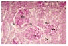
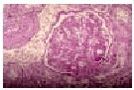
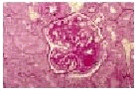
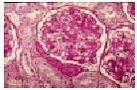
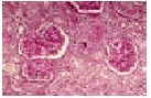
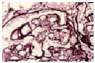
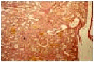
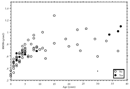
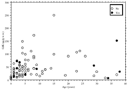
6 Effects6.1 Cadmium toxicity to ringed seals6.2 Effects of contaminants in the Greenland Sea Polar bear 6.3 References Citation: C. Sonne-Hansen1, R. Dietz,1 F. Riget1, E. Born2, L. Hyldstrup3& P. S. Leifsson4 Chapter 6. Effects. In: Riget, F., J. Christensen and P. Johansen (eds). AMAP, Greenland and the Faroe Islands 1997-2001. Ministry of Environment, Denmark 6.1 Cadmium toxicity to ringed sealsA total of 100 ringed seals from 1998 sampled in Qaanaaq, North West Greenland were examined to detect cadmium induced nephropathy and osteodystrophy. Cadmium concentrations in the kidneys were determined and histological examination of the kidneys and DXA scanning of the sceletal system (lumbal vertebraes) were conducted as described in Sonne-Hansen et al. (2000, 2002). The background of the study is that the Greenland marine food chains contain high levels of cadmium, mercury and selenium. Concentrations of cadmium in the kidney of ringed seals from the municipalities of Qaanaaq and Upernavik (northwest Greenland) are among the highest recorded in the Arctic and therefore selected for the study. The purpose of the study was to determine whether cadmium induced damage in the kidneys and the skeletal system could be detected among 100 ringed seals from Northwest Greenland. The cadmium concentrations in the kidney cortex ranged from 0 to 248 µg/g ww (mean = 44.5 µg/g ww) in the 99 kidneys examined. Experience from cadmium poisoned humans and laboratory mammals indicate that concentrations above 50-200 µg/g ww may induce histopathological changes (Friberg, 1986; WHO, 1992; Elinder & Järup, 1996). Overall, 31 of the ringed seals had cadmium concentrations in the kidney cortex above 50 µg/g ww, 11 had concentrations above 100 and 1 had concentrations above 200 µg/g ww (see Figure 6.1.9). Obvious histopathological changes (categorized mainly as glomerulonephritis) were found in 10 of the seals, however, none of these changes could specifically be atributed to cadmium induced renal damage (mainly tobulopathy) as described for other species (see Table 6.1.1 and Figure 6.1.7). Damage to the proximal kidney tubules is known to induce demineralisation of the skeletal system (Fanconi’s syndrom). Therefore the three lowest lumbar vertebrae were scanned in 91 seals to measure the content of calcium. The 10 cases of nephropathy could neither be linked to the degree of mineralisation of the skeleton or to the cadmium concentrations (see Figure 6.1.8 and 6.1.9). Furthermore, the degree of mineralisation of the skeleton was not correlated with the cadmium concentration or sex (p=0.68), but was highly significant correlated to age (p<0.001).
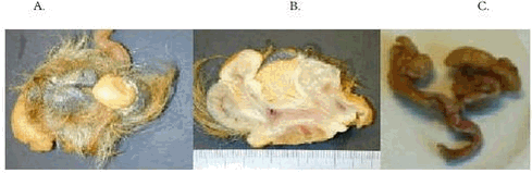
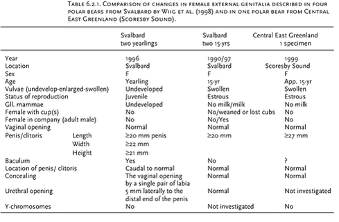
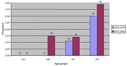
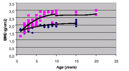
It can be concluded that 10% of the ringed seals showed renale histopathological changes. Despite high levels of cadmium it cannot be concluded whether the ringed seals showed cadmium-induced nephropathy or osteodystrophy. This might be explained by the composition of the ringed seals diet, which contains high levels of vitamin D, calcium, phosphorus, zinc, selenium and protein. These elements are all likely to counteract cadmium-induced damage. It is speculated that ringed seal are not particularly vulnerable to osteodystrophy, due to their continuous growth (bone mineralisation) throughout life and the female estrogen hormonal activity throughout life. 6.2 Effects of contaminants in the Greenland Sea Polar bearRecently, there has been an increased focus on the negative effects on Arctic organisms of organochlorines (OCs) that originate in the industrialized parts of the world. Long-lived and persistent substances such as DDT and PCB become concentrated in food webs, from lower to higher trophic levels. As apex predator in the Arctic marine ecosystem, polar bears acquire relatively large burdens of OCs because their diet consists primarily of seal blubber and meat. Bears in Svalbard, the western Russian Arctic and East Greenland have relatively high concentrations of OCs, and their levels are high enough to raise concern about effects on health and reproductive performance. Propably 17 cases of female polar bears with deformities in their external sex organs (pseudohermaphrodites) have been recorded at Svalbard since 1990 (Derocher pers. comm.). These deformities are suspected of being caused by high levels of OCs (Wiig et al. 1998). In 1999 the NERI and the Greenland Institute of Natural Resources (Nuuk) initiated a study to assess the effects of OCs on internal and external organs of polar bears in east Greenland. The study involves: (1) obtaining information from the Greenland polar bear hunters concerning their observations of bears with aberrant organs or behavior, The project is ongoing and still awaits the ageing of the individuals, results from additional chemical analysis and histological examinations. Therefore, the results presented below are regarded as preliminary and no confirm conclusions should be made. Aberrant polar bears in east Greenland Dietz et al. (2001) investigated the frequence of macroscopic pathological changes detected by local hunters - in polar bears in east Greenland in 1999. 52 local east Greenland hunters from Scoresby Sound and Ammassalik were interviewed and they covered at least 1110 polar bears in total. Despite the female polar bear with mega-clitoris 12 bears with aberrant macroscopic divergation were reported of which 8 could be congenital maldeformation and/or due to other aetiology like endocrine disruption. The changes were malformation of a new born, papillae mammae accessoria, 2 bears with six claws, one bear with missing claws on left and right hind paws and 3 bear with diverging degrees of focal macular melanoses (due to inflammation and/or formation of connective tissue). This is a 0.81% (9/1110) frequency of aberrant polar bears due to environmental estrogens and/or with other aetiology which is lower than expected compared to e.g. frequencies in populations in domestic and wild animals. The frequency of pseudohermaphroditism among east Greenland polar bears are 0.09% (1/1110) and lower than the frequency of for example female pseudohermaphrodites oberserved at Svalbard (2-3%) (Wiig et al. 1998, Derocher pers. comm.). Female pesudohermaphrodite polar bears in east Greenland and at Svalbard The female pseudohermaphrodite from east Greenland were investigated by veterinarian-pathologists in March 2002 and macroscopically both the internal and external reproductive organs were normoanatomical and -functional except for the megaclitoris (Figure 6.2.1). Compared to samples obtained from other female polar bears the reproductive organs are normal in anatomy and functionality (see Table 6.2.1). The female polar bear pseudohermaphrodite was in estrus with an active ovarium without a dominating follicle (>10 follicles in each ovarium and a corpus luteum). Genetically the female polar bear were a female with no chormosomal abnormalities or presence of a y-chromosome (Andersen pers. comm.). At Svalbard 4 polar bear female pseudohermaphrodites have been reported from 1990-97 (Wiig et al. 1998). Afterwards app. 13 suspected female polar bear pseudohermaphrodites has been clinically examined during helicopter survey and radiotelemetry investigations but these are not reported yet (Derocher pers. comm). The east Greenland bear has been examined post mortem after formaldehyde-alcohol fixation of reproductive organs and endocrine glands. Table 6.2.1 summarises the results from the clinically macroscopic examinations of the external genitalia of the bears. Female pseudohermaphroditism is known to be induced by endocrine disruption foetally or post partum (Benirschke 1981, Polani 1981, Capen 1995, Feldman 1995, Mickelsen & Menon 1995, Wiig et al. 1998). Four hypotheses have been mentioned (Ibid.): (1) Freemartiinism (when the male of two twins influence the female foetus by its androgens through the anasotmoses in the foetal vascular system). (2) Tumors (the mother has produced more testosterone than normal due to a tumor in the brain or the ovaries which gives the female foetus male characteristics). (3) Mutations (genetic mutations where pseudohermaphroditism becomes congenital). (4) POPs (persistent organic pollutants where anthropogenic xenoandrogens and/or estrogens disrups the endocrine homeostasis in the mother and the foetus are malformed due to altered hormonal status). The female pseudohermaphrodite polar bears from Svalbard could hence reflect a congenital pseudohermaphrodite complex, an androgenproducing tumor or POP pollution but pobably not due to freemartiinism (Benirschke 1981). The east Greenland female pseudohermaphrodite polar bear - as it is the single one observed in this study and by local hunters – is likely to be caused by an androgenproducing tumor, androgensubstances (OCs) or be a polar bear from Svalbard. Fluctuating asymmetry and osteopenia and macroscopic skull pathology in east Greenland polar bears A total of 283 polar bear skulls were investigated to detect fluctuating asymmetry and macroscopic pathology (parodontitis) over time. Fluctuating asymmetry has been used as a stress indicator, in numerous animal and plant organisms in wildlife and laboratory mammals, to reflect developmental instability (e.g. Palmer & Strobeck 1986, Nachman & Heller 1999). To detect the degree of fluctuating asymmetri 16 metric bilaterally traits and 17 meristric as foraminas (canal openings in the skull for nerves and blood vessels) were examined in the skulls covering the time period from 1892 to 2000. In a preliminary analyses, the skulls were divided into two groups; before and after 1960 and it was tested whether fluctuating asymmetry was occuring and whether there was differences between the two peiod. The analyses was preliminary because the skulls could not be divided according to the age of the bears. The analysis gave no indications of the skulls from after 1970 (the OC pollutants affected period) to have higher degree of fluctuating asymmetry than skulls before 1970. The frequency of parodontitis within preliminary age groups was also investigated in skulls before and after 1970 (Figure 6.2.2). The frequency of parodontitis increased significantly at the 5% level with age and no differences was found between sampling periods (logistic regression analysis). A X-ray bone densitometer was used to determine the mineralisation of the skulls (calcium-phospate content). The data were analysed using a software programme, which generates a picture of the bone segment and calculates the bone mineral content (BMD, g/cm2). Figure 6.2.3 shows the relationship between BMD and the preliminary age, indicating lower BMD in females than in males. This may be expected as female mobilises calcium-phosphate to the foetus and later on also to the weaning pub.
6.3 ReferencesBenirschke, K., 1981. Hermaphrodites, freemartins, mosaics and chimaeras in animals. In: C. R. Austin and R. G. Edwards (eds.): Mechanisms of sex differentiation in animals and man (Vol. II). Academic Press, London, UK: 421-463. Capen, C.C., 1995.Endocrine system. In: Carlton, W.W. & M.D. McGavin (eds.): Thomson’s special veterinary pathology (2nd edn.). Mosby-Year Book, Inc., St. Louis, USA: 247-284. Dietz, R., C. Sonne-Hansen, E.W. Born, H. Sandell & B. Sandell, 2001. Aberrant polar bears in East Greenland: An interview investigation. National Environmental Research Institute, Denmark: Technical Report No. 359: 51 pp. Elinder, C.-G., & L. Järup, 1996. Cadmium and health risks: Recent findings. Ambio 5: 370-373 Feldman, E. C., 1995. Hyperadrenocorticism. In: Ettinger, S. J. and E. C. Feldman (eds.): Textbook of veterinary internal medicine (vol. II). W.B. Saunders Company, Philadelphia, USA: pp. 1538-1578. Friberg, L., C.-G. Elinder, T. Kjellström, and G.F. Nordberg (1986): Cadmium and Health. A toxicological and epidemiological appraisal volume II. CRC Press, Inc., Boca Raton, Florida, USA: 303 pp. Mickelsen, W.D. & M.A. Memon, 1995.-The reproductive system – Inherited and congenital disorders of the male and female reproductive systems. In: Ettinger, S.J. & E.C. Feldman (eds.): Textbook of veterinary internal medicine (vol. II). W.B. Saunders Company, Philadelphia, USA: 1686-1689. Nachman, G. & K. E. Heller, 1999. Fluctuating asymmetry as an index of fitness: causality or statistical artifact? Oikos 86 (2): 357-365. Palmer, A. R. & C. Strobeck, 1986. Fluctuating asymmetry: measurement, analysis and pattern. Annu. Rev. Ecol. Syst. 17: 391-421. Polani, P. E., 1981. Abnormal sex development in man. Anomalies of sex-differentiating mechanisms (II). In: C. R. Austin and R. G. Edwards (eds.): Mechanisms of sex differentiation in animals and man (Vol. II). Academic Press, London, UK: 467-547. Sandell, H.T. , B. Sandell, E.W. Born, R. Dietz & C. Sonne-Hansen, 2001.-Isbjørne i Østgrønland. En interviewundersøgelse om forekomst og fangst, 1999. Teknisk Rapport nr. 40. Pinngortitaleriffik, Grønlands Naturinstitut, Nuuk: 96 pp. Sonne-Hansen C., R. Dietz, P. S. Leifsson, L. Hyldstrup & F. F. Riget, 2000. Cadmium toxicity to ringed seals (Phoca hispida). An epidemiological study of possible cadmium induced nephropathy and osteodystrophy in ringed seals (Phoca hispida) from Qaanaaq in Northwest Greenland. National Environmental Research Institute, Denmark. NERI Technical Report No. 307, 31 pp. Sonne-Hansen, C., R. Dietz, P. S. Leifsson, L. Hyldstrup & F. F. Riget, 2002. Cadmium toxicity to ringed seals (Phoca hispida) - An epidemiological study of possible cadmium induced nephropathy and osteodystrophy in ringed seals (Phoca hispida) from Qaanaaq in Northwest Greenland. Sci. Tot. Env. 295 (I-III): 167-181. WHO, 1992. IPCS - Environmental Health Criteria 134: Cadmium. WHO, Geneva, Schweiz: pp. 1-209. Wiig, Ø., A.E. Derocher, M.M. Cronin & J.U. Skaare, 1998.-Female pseudoherma-phrodite polar bears at Svalbard. J. of Wildl. Diseases, 34(4): 792-796. |