Effects of azole fungicides on the function of sex and thyroid hormones
3 Results - Endocrine disrupting effects of azole fungicides
- 3.1 In vitro effects
- 3.1.1 Estrogenic/anti-estrogenic effects of azole fungicides
- 3.1.2 Aromatase effects of azole fungicides
- 3.1.3 Anti-androgenic effects of azole fungicides
- 3.1.4 Dioxin-like effects of azole fungicides – Ah-receptor testing
- 3.1.5 Steroid synthesis effects of azole fungicides
- 3.1.6 Thyroid effects of azole fungicides
- 3.2 In vivo effects
- 3.3 Overview of results
3.1 In vitro effects
To investigate the endocrine disrupting mechanisms involved, the four azole fungicides: epoxiconazole, prochloraz, propiconazole, and tebuconazole were tested in a range of in vitro test systems to evaluate anti/estrogenicity, anti/androgenicity, dioxin like effects, disruption of the thyroid function, and effects on aromatase activity and other enzymes involved in the steroid synthesis.
3.1.1 Estrogenic/anti-estrogenic effects of azole fungicides
Figure 4 illustrates that all four fungicides inhibited MCF-7 cell proliferation induced by 10 pM 17β-estradiol and hence exhibited weak anti-estrogenic responses. The response was statistically significant from 1.6 µM (Table 3). Concentrations causing 25%, 50%, and 75% inhibition of the 17β-estradiol induced (10 pM) MCF-7 cell proliferation response (IC25 , IC50, IC75) were calculated (Table 3). IC50 values were found to be 49, 28, 52, and 45 µM for epoxiconazole, prochloraz, propiconazole, and tebuconazole, respectively. Prochloraz was the most potent of the four fungicides.
Table 3 - Concentrations (µM) of the fungicides causing 25, 50, or 75 % inhibition of the 17β-estradiol (10 pM) induced MCF-7 cell proliferation response.
Epoxiconazole Prochloraz Propiconazole Tebuconazole
LOEC 25 12.5 25 1.6
IC25 36 21 40 32
IC50 49 28 52 45
IC75 66 38 64 63
LOEC: Lowest observed effect concentration. LOEC is chosen as the lowest concentration causing a continuously statistically significant response.
Since a decreased proliferation can be due to general cell toxicity of the fungicides in the MCF-7 cells, a LDH-test for cytotoxicity was performed. Cytotoxicity was only detected at the highest azole concentrations (indicated by a c in the figures) and cannot explain the decreased cell proliferation of the azole fungicides at lower concentrations.
In addition, epoxiconazole and propiconazole increased the cell proliferation indicating a weak estrogenic activity (Figure 5). This activity was statistically significant at 6.25 and 25 µM, respectively.
Experiments with the well-known anti-estrogenic compound ICI 182.780 (1 nM) revealed that the cell proliferation induced by these fungicides was counteracted by ICI 182.780, thus, indicating that the proliferation is induced directly via the ER receptor (data not shown).
Figure 4 - Anti-estrogenic effects of the four fungicides: epoxiconazole, prochloraz, propiconazole, and tebuconazole tested in the MCF-7 cell proliferation assay. All four fungicides inhibited the proliferation of MCF-7 cells induced by 10 pM 17β-estradiol. Data represent mean±SD for three to seven independent experiments. The horizontal gray line represents the control level and the dashed gray lines represent the SD of the control. * Statistically significantly different from control (10 pM 17β-estradiol) (p<0.05). c = tested cytotoxic by use of a LDH-test. RPE = Relative Proliferative Effect.
Figure 5 - Estrogenic effects of the four fungicides: epoxiconazole, prochloraz, propiconazole, and tebuconazole tested in the MCF-7 cell proliferation assay. Epoxiconazole and propiconazole induced proliferation of the MCF-7 cells. Data represent mean±SD for three to seven independent experiments. The horizontal gray line represents the control level and the dashed gray lines represent the SD of the control. * Statistically significantly different from control (p<0.05). c = tested cytotoxic by a LDH-test. RPE = Relative Proliferative Effect.
3.1.2 Aromatase effects of azole fungicides
Figure 6 illustrates that all four fungicides inhibit the 1µM testosterone-induced MCF-7 cell proliferation. The effect is statistically significant from 1 µM. The response obtained in this assay will be the combined effect of aromatase inhibition and anti-estrogenicity as aromatase converts testosterone to estrogen, which then induces proliferation of the cells.
Concentrations causing 25%, 50%, and 75% inhibition of the 1 µM testosterone induced MCF-7 cell proliferation response (IC25, IC50, IC75) were calculated (Table 4). IC50 values were found to be 17, 9, 29, and 19 µM for epoxiconazole, prochloraz, propiconazole, and tebuconazole, respectively. Prochloraz was the most potent among the four (Table 4).
The concentrations of the azole fungicides needed to reduce the testosterone-induced response were lower than the concentrations needed to reduce the 17β-estradiol-induced response. This indicates that these compounds possess both aromatase inhibiting and anti-estrogenic properties but that aromatase inhibition dominates at low concentrations.
Figure 6 - Effects on the enzyme aromatase of the four fungicides: epoxiconazole, prochloraz, propiconazole, and tebuconazole tested in the MCF-7 cell proliferation assay. All four fungicides inhibited the 1 µM testosterone (converted by aromatase to estrogen) induced MCF-7 cell proliferation. Data represent mean±SD for three independent experiments. The horizontal gray line represents the control level and the dashed gray lines represent the sd of the control. *Statistically significantly different from control (1 µM testosterone) (p<0.05). c = tested cytotoxic by a LDH-test. RPE = Relative Proliferative Effect.
Table 4 - Concentrations (µM) of the fungicides causing 25, 50, or 75 % inhibition of the testosterone-induced (1 µM) MCF-7 cell proliferation response.
Epoxiconazole Prochloraz Propiconazole Tebuconazole
LOEC 1 1 10 10
IC25 4 3 16 6
IC50 17 9 29 19
IC75 73 25 53 60
LOEC: Lowest observed effect concentration. LOEC is chosen as the lowest concentration causing a continuously statistically significant response.
3.1.3 Anti-androgenic effects of azole fungicides
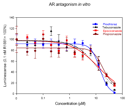
All four azole fungicides proved to be AR antagonists. No cytotoxicity was observed except for prochloraz and tebuconazole that were cytotoxic at 50 μM (data not shown). As shown in Figure 7 all the azole fungicides had a potency of comparable magnitude. The LOECs were 0.8, 3.1, 25 and 3.1 μM for epoxiconazole, prochloraz, propiconazole, and tebuconazole respectively.

Figure 7 - Anti-androgenic effects determined in the AR reporter gene assay. The fungicides are tested in combination with the AR agonist R1881 in AR transfected CHO cells. Data represent mean±SD for three independent experiments. * Significance level p < 0.05 compared to the control (0.1 nM R1881).
3.1.4 Dioxin-like effects of azole fungicides – Ah-receptor testing
Like prochloraz (Long et al., 2003) epoxiconazole, propiconazole, and tebuconazole were able to activate the Ah receptor (Figure 8), but far from the order of magnitude displayed by prochloraz. The LOECs were 6.3, 0.05, 12.5 and 6.3 μM for epoxiconazole, prochloraz, propiconazole, and tebuconazole, respectively (Table 5).
Table 5 - Concentrations (mM) of the fungicides needed to induce a significant response in the AhR CALUX reporter gene assay, and the effect of the fungicides in relation to the maximum response induced by the AhR agonist TCDD.
Epoxiconazole Prochloraz Propiconazole Tebuconazole
LOEC 6.3 0.05 12.5 6.3
MOEC 50 10 50 50
% Effect of
max.
TCDD effect 9 38 10 8
LOEC: Lowest observed effect concentration. LOEC is chosen as the lowest concentration causing a continuously statistically significant response. MOEC: Maximum observed effect concentration: the lowest concentration causing maximum response.
Figure 8 - Agonistic effect measured in the AhR CALUX reporter gene assay. Concentration response curves for the four fungicides epoxiconazole, prochloraz, propiconazole, and tebuconazole and for the AhR agonist TCDD. For concentrations above 10 μM a decrease of luciferase activity was seen for prochloraz, which is due to a cytotoxic effect. * The prochloraz data is from a previous conducted study in 2002.
3.1.5 Steroid synthesis effects of azole fungicides
In order to look for effects on steroidogenesis in vitro, we tested epoxiconazole, prochloraz, propiconazole and tebuconazole in the H295R steroid synthesis assay, and compared it to the results of the previously conducted study with prochloraz (Laier et al., 2006).
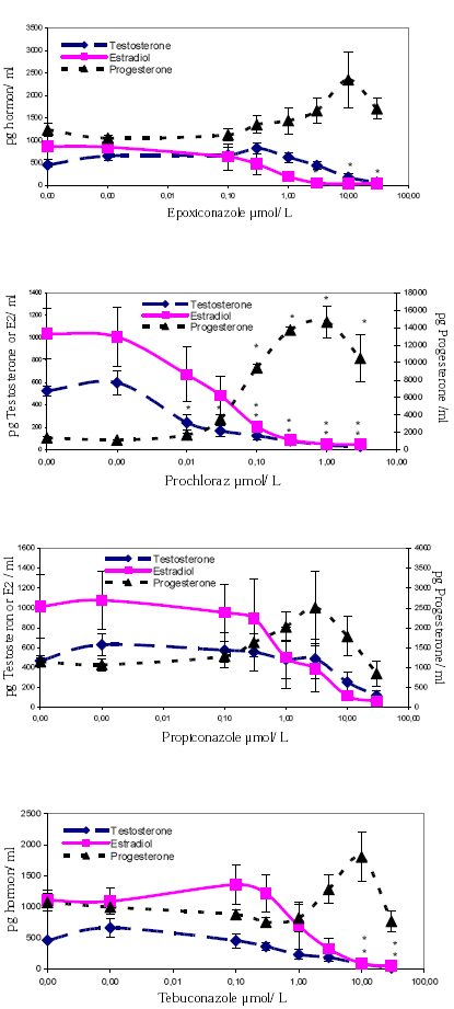
Epoxiconazole, propiconazole, and tebuconazole, were like prochloraz able to inhibit the production of testosterone and estradiol in vitro in H295R cells, though the inhibiting effect on testosterone production was only statistically significant for the two highest concentrations of tebuconazole. The estradiol production was statistically significantly inhibited at the two highest concentrations of tebuconazole, and the tree highest concentrations of epoxiconazole (Figure 9). Regarding effects on progesterone synthesis, the general picture was a stimulating effect, but at the highest concentrations of all fungicides the stimulation was decreased (Figure 9). Cytotoxicity was not found at any concentrations using the resazurin-test.

Figure 9 - In vitro effects of the fungicides on testosterone, progesterone and estradiol formation in human adrenocortical carcinoma cells (H295R). Data represent the mean±SEM for two to three independent experiments. E2 = estradiol. * Statistically significantly different from control (P<0.05).
3.1.6 Thyroid effects of azole fungicides
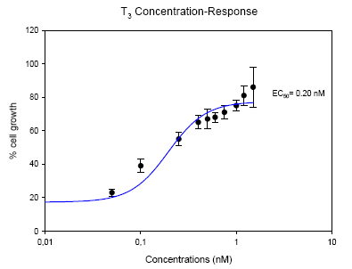
The effect on proliferation of GH3 rat pituitary tumor cells in vitro by T3 and the fungicides is illustrated in Figure 10 and 11, respectively. T3 dose-dependently stimulated the cell proliferation at 0.05 nM (LOEC) and maximally at 1.5 nM (MOEC) (Figure 10). The half maximum response (EC50) of T3 was determined at 0.20 nM. This assay was established as part of this project and the fact that we get a nice dose-response curve for T3 verifies that the assay works.

Figure 10 - Effect of T3 on proliferation of GH3 cells. Solvent control (0.1% DMSO) was set to 0 %.
EC50 determined to 0.20 nM. Data represent mean±SD for three independent experiments.
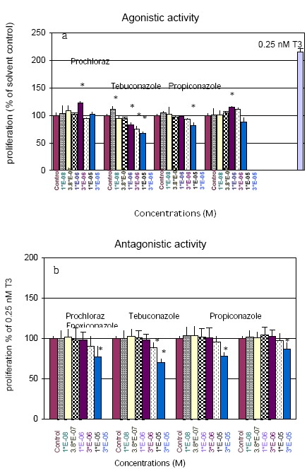
Propiconazole and tebuconazole had a weak inhibitory effect on GH3 cell growth (Figure 11). At concentrations of 3 µM to 30 µM tebuconazole statistically significantly decreased the cell proliferation to approximately 70% of basal level and propiconazole significantly decreased the cell proliferation at 30 µM to approximately 80% of solvent control.
Tebuconazole significantly inhibited the T3-induced growth of the GH3 cells at 10 and 30 µM, and for the three other pesticides a significant decreased cell growth was also seen at 30 µM (Figure 11). The decrease in cell proliferation at 30 µM, for cells treated with the fungicides alone as well as upon co-treatment 0.25 nM T3, is probably due to a cytotoxic effect, as others have demonstrated cytoxicity at these concentrations, for some of the same chemicals (Ghisari and Bonefeld-Jorgensen, 2005).

Figure 11 - Effects of the fungicides in the T-screen assay: GH3 cells treated with different concentrations of fungicides alone (a) or in the presence of 0.25 nM T3 (b). The values are given as percentage of the proliferation of solvent control (0.1% DMSO) or of positive control (0.25 nM T3). Data represent mean±SD. * Statistically significantly different (p £ 0.05 ) from the respective controls.
3.2 In vivo effects
A Hershberger test and a developmental toxicity study were conducted to investigate in vivo effects of the azole fungicides.
3.2.1 Anti-androgenic effects in the Hershberger test
Propiconazole and tebuconazole were tested in castrated testosterone-treated rats in order to determine if the compounds were able to block androgen receptors or affect androgen levels in vivo. They were administered orally at doses of 50, 100, and 150 mg/kg together with a sc injection of a fixed dose of testosterone. The anti-androgen flutamide was given as a positive control.
In vivo, body weights, paired kidney weights, thyroid and pituitary weights were unaffected by the treatments, whereas liver weights were increased with both fungicides. The increase in liver weights was statistically significant at the highest dose of tebuconazole and the two highest doses of propiconazole.
Compared to intact males of the same age, castrated vehicle-exposed rats had significantly reduced weights of all the reproductive organs and increased levels of LH and FSH (Table 6 and 7). The serum levels of T4 were unaffected by castration. On the gene expression level both PBP C3 and ODC mRNA were reduced by castration, whereas TRPM-2 and Compl. C3 mRNA were increased (Table 8). All these results were in accordance with predictions based on theoretical knowledge and previous experiments.
Flutamide exerted the expected effects as well, and caused qualitatively the same effects as seen after castration. The weights of all reproductive organs were decreased; serum LH and FSH levels were increased and at gene level both PBP C3 and ODC mRNA were reduced, whereas TRPM-2 and Compl. C3 mRNA were increased. Overall it can be concluded that this Hershberger test was performed as expected.
The fungicides had no effect on neither reproductive organ weights or on hormone levels, except for the highest dose of propiconazole, for which there was a significant increase in FSH. On the gene expression level, the only significant effect of the fungicides was a decrease in the level of ODC mRNA in prostate, with all doses of propiconazole and with the highest dose of tebuconazole. This gene is regulated by androgens but is also regulated via other pathways, so in the light of the lack of anti-androgenic effects on other genes, it is conceivable that this effect is due to a non-androgen dependent effect.
In conclusion neither propiconazole nor tebuconazole had any androgen receptor blocking effects in vivo in the Hershberger test at doses below 150 mg/kg bw/day. This is in contrast to the imidazole fungicide prochloraz that induced anti-androgenic effects at doses between 50 and 150 mg/kg bw/day.
Table 6 - Body and Organ weights of young male rats in the Hershberger test.
Click here to see the Table.Table 7 - Serum hormone levels of young male rats in the Hershberger test.
Click here to see the Table.Table 8 - Gene expression in ventral prostate of young male rats in the Hershberger test.
Click here to see the Table.3.2.2 Effects on offspring after prenatal exposure
Developmental effects of epoxiconazole and tebuconazole were investigated by dosing pregnant dams with test compounds and examination of their fetuses and offspring for effects on sexual differentiation.
3.2.2.1 Effects on GD 21 fetuses
As seen from Table 9 the highest dose of tebuconazole lead to a decreased body weight gain in dams, probably due to effects on both the dam and the uterine content, increased loss of fetuses and decreased fetal weight on GD 21. The highest dose of epoxiconazole caused increased loss of fetuses and decreased fetal weight in the absence of effects on maternal weight gain. Many of the dead fetuses (27 of 128) had died very late in the gestation period, while such late fetal death was not seen in the controls (0 of 70). The lower dose of epoxiconazole also seemed to induce increased fetal death, but the differences were not statistically significant. Very late fetal death occurred in 4 out of 92 cases compared to 0 in the controls. Although not statistically significant these findings raise concern for effects on fetal survival also at this dose level.
3.2.2.2 Immunohistochemistry
StAR and P450scc immunostaining were comparable to controls. However, it cannot be excluded that effects on PBR and 17β HSD immunostaining were present, but due to lack of control foetuses for this analysis, no conclusions could be made.
Table 9 - Pregnancy and litter data for the developmental study.
| Control | Tebuconazole 50 mg/kg |
Tebuconazole 100 mg/kg |
Epoxiconazole 15 mg/kg |
Epoxiconazole 50 mg/kg |
|
| Dams and litters | |||||
| No. of dams (viable litters) | N=13 (13) | N=12 (12) | N=10 (8) ¤ | N=10 (9) | N=7 (1-2) ¤ |
| BW gain. GD7-GD21 (g) | 85.38 ± 11.9 | 77.17 ± 14.4 | 61.00±12.5* | 87.70 ± 16.1 | 92.14 ± 14.0 |
| BW gain. GD7-PND1 (g) | 20.62 ± 7.2 | 17.58 ± 6.8 | 13.50 ± 10.6* | 18.11 ± 6.7 | 15.00 ± 8.5 |
| BW gain PND1-PND13 (g) | 7.53 ± 16.3 | 7.75 ± 16.84 | -4.57 ± 12.9 | -6.89 ± 24.8 | 20 |
| Gestation length (d) | 22.46 ± 0.5 | 22.67 ± 0.5 | 23.40 ± 1.2** | 22.67 ± 0.7 | 23.71 ± 0.8** |
| % postimplantation loss | 6.55 ± 5.1 | 10.31 ± 11.2 | 27.32 ± 23.5* | 16.01 ± 30.0 | 34.25 ± 18.2* |
| % perinatal loss | 9.67 ± 8.0 | 13.37 ± 12.5 | 54.97 ± 36.9** | 18.21 ± 30.0 | 88.78 ± 29.7** |
| Litter size | 11.15 ± 1.7 | 10.75 ± 3.6 | 8.75 ± 3.8 | 9.90 ± 4.1 | 8.67 ± 3.1 |
| Born alive per litter | 10.92 ± 1.7 | 10.67 ± 3.7 | 8.38 ± 3.8 | 9.80 ± 4.0 | 4.33 ± 5.9** |
| Born dead per litter | 0.23 ± 0.4 | 0.08 ± 0.3 | 0.37 ± 0.7 | 0.10 ± 0.3 | 4.33 ± 2.9** |
| % PN death | 3.39 ± 5.55 | 3.36 ± 7.04 | 27.00 ± 37.5* | 2.78 ± 5.9 | 69.44 ± 52.9** |
| % males | 44.76 ±17.6 | 56.42 ± 11.7 | 40.36 ± 18.6 | 45.45 ± 13.13 | 34.72 ± 33.42 |
| Offspring (data from viable litters) | |||||
| Birth weight (g) | 5.53 ± 0.3 | 5.64 ± 0.5 | 5.63 ± 0.8 | 6.21 ± 0.6** | 6.36 ± 0.3 |
| BW. PND 13 (g) | 23.25 ± 2.6 | 21.59 ± 4.14 | 22.39 ± 5.02 | 21.48 ± 4.8 | 23.4 |
| Male AGD (mm) | 3.41 ± 0.2 | 3.39 ± 0.1 | 3.51 ± 0.2 | 3.65 ± 0.22* | 3.41 ± 0.3 |
| Male AGD/cuberoot bw | 1.92 ± 0.1 | 1.90 ± 0.1 | 1.96 ± 0.1 | 1.96 ± 0.1 | 1.83 ± 0.2 |
| Female AGD (mm) | 1.72 ± 0.1 | 1.80 ± 0.1 | 1.91 ± 0.1* | 1.95 ± 0.2** | 1.71 |
| Female AGD/cuberoot bw | 0.98 ± 0.03 | 1.02 ± 0.06 | 1.09 ± 0.07* | 1.08 ± 0.08* | 0.96 |
| No. aerolas in males | 2.08 ± 0.6 | 3.43 ± 0.9** | 3.07 ± 2.5** | 2.53 ± 1.1 | 3.38 |
| No. aerolas in females | 12.5 ± 0.4 | 12.46 ± 0.4 | 12.31 ± 0.4 | 12.32 ± 0.2 | 12 |
| GD21 Caesarian section ¤ | |||||
| No. of dams | N=6 | N=7 | N=8+2 ¤ | N=9 | N=14+4 ¤ |
| Maternal bw (g) | 307.17 ± 22.4 | 297.00 ± 27.2 | 281.00 ± 26.5* | 287.33 ± 29.8 | 285.73 ± 17.7 |
| Adjusted bw (g) | 232.70 ± 14.9 | 234.00 ± 16.8 | 223.31 ± 20.4 | 231.49 ± 15.9 | 223.13 ± 21.0 |
| No. implantations | 12.50 ± 2.1 | 12.00 ± 3.2 | 11.60 ± 1.5 | 10.22 ± 4.3 | 12.06 ± 2.4 |
| No. fetuses | 11.67 ± 2.1 | 11.14 ± 3.6 | 9.40 ± 2.1 | 9.11 ± 4.9 | 9.00 ± 4.1 |
| % postimplantation loss | 6.45 ± 7.9 | 9.10 ± 11.3 | 21.54 ± 9.4* | 20.87 ± 33.5 | 28.14 ± 26.9* |
| % late res | 1.28 ± 3.1 | 2.38 ± 6.3 | 6.14 ± 4.2 | 13.89 ± 33.3 | 24.88 ± 27.3* |
| % very late res | 0.0 ± 0.0 | 2.38 ± 6.3 | 2.39 ± 4.2 | 4.16 ± 11.8 | 15.12 ± 24.0* |
| % males | 56.01 ± 17.2 | 46.22 ± 20.9 | 49.74 ± 20.6 | 46.41 ± 18.8 | 53.57 ± 25.3 |
| Fetal weight male (g) | 4.45 ± 0.3 | 3.84 ± 0.7 | 3.44 ± 0.9** | 3.98 ± 0.9 | 3.79 ± 0.7* |
| Fetal weight female (g) | 4.18 ± 0.4 | 3.61 ± 0.6 | 3.40 ± 0.9* | 3.83 ± 0.8 | 3.54 ± 0.7* |
| No. litters for AGD # | N=3 | N=4 | N=4 | N=6 | N=10 |
| Male AGD (mm) | 3.39 ± 0.3 | 3.50 ± 0.03 | 3.29 ± 0.4 | 3.54 ± 0.1 | 3.40 ± 0.1 |
| Male AGD /cuberoot bw | 2.08 ± 0.1 | 2.25 ± 0.2 | 2.30 ± 0.1* | 2.31 ± 0.1* | 2.25 ± 0.1 |
| Female AGD (mm) | 1.65 ± 0.1 | 1.87 ± 0.2* | 2.02 ± 0.1** | 1.91 ± 0.3** | 1.92 ± 0.1** |
| Female AGD/cuberoot bw | 1.04 ± 0.1 | 1.23 ± 0.2 | 1.43 ± 0.2** | 1.28 ± 0.2* | 1.29 ± 0.1** |
Data represent group means based on litter means±SD.
PN = postnatal. Res = resorption i.e. regression of the fetus;
* or ** statistically significant compared to control (p<0.05 or p<0.01, respectively). Significant values are indicated in bold.
¤ Because of problems with parturition caesarian section (CS)(GD23-25) was performed on 2 dams in the 100 mg/kg tebuconazole group and 4 dams in the 50 mg/kg epoxiconazole group. These data were included in the analysis of GD21 CS data.
# AGDs were only measured in the 2nd set of animals.
Values written in Italic are from only one dam/litter why no SD are shown.
3.2.2.3 Hormone analysis
In fetuses tebuconazole caused a statistically significant increase in testicular 17a-hydroxy-progesterone and progesterone levels, and a decrease in testosterone levels. Epoxiconazole had no significant effect on the measured hormone levels in fetuses (Table 10).
Table 10 - Testicular hormone concentrations in male fetuses at GD21.
| 17α-hydroxy- progesterone (pg/testis) |
Testosterone (ng/testis) |
Progesterone (ng/testis) |
Testosterone production (ng/testis) |
Progesterone production (ng/testis) |
|
| Control | 1.95±0.54(4) | 1.75±0.71(5) | 0.037±0.025(5) | 3.95±1.71(6) | 0.02±0.01(6) |
| Tebuconazole 50 mg/kg | 8.39±2.59*7) | 1.25±0.40(7) | 0.103±0.035* (7) | 4.77±3.49(6) | 0.29±0.62(6) |
| Tebuconazole 100 mg/kg | 6.59±3.88* (9) | 0.88±0.46* (9) | 0.084±0.063(9) | 3.34±2.59(5) | 0.04±0.03(5) |
| Epoxiconazole 15 mg/kg | 1.76±1.36(6) | 1.62±0.59(8) | 0.029±0.019(8) | 4.41±3.27(6) | 0.00±0.00(6) |
| Epoxiconazole 50 mg/kg | 0.94±0.48 (13) |
1.11±0.56 (20) |
0.027±0.019 (20) |
4.00±3.27 (10) |
0.00±0.01 (10) |
Fetal testes were extracted with diethylether before measurement of progesterone, 17α-progesterone and testosterone. Other fetal testes were incubated in a water bath at 37°C for 3 hrs before measurement of testosterone and progesterone in supernatants. Data represent the mean±SD; ( ) = n; * statistically significantly different compared to control (P<0.05).
In plasma from mothers obtained at GD 21, the highest dose of tebuconazole led to a marked increase in the progesterone level (7-fold increase), and a significant decrease in T3. Epoxiconazole led to an increase in the progesterone (7-fold increase) and surprisingly also to a pronounced increase in the testosterone level (2-fold), at the highest dose (Table 11). The increased progesterone levels in the mothers could be the explanation for the increased gestational length (Table 9).
Table 11 - Maternal plasma concentrations of hormones and lipids at GD 21.
Click here to see the Table.3.2.2.4 Gene expression levels
Table 12 - Overview of gene expression in testis from male fetuses (GD 21).
| Control | Tebuconazole 50 mg/kg |
Tebuconazole 100 mg/kg |
Epoxiconazole 15 mg/kg |
Epoxiconazole 50 mg/kg |
|
| ScarB1 mRNA | ↔ | ↔ | ↔ | ↔ | ↔ |
| P450scc mRNA | ↔ | ↔ | ↔ | ↔ | ↔ |
| P450c17 mRNA | ↔ | ↔ | ↔ | ↔ | ↔ |
| StAR mRNA | ↔ | ↔ | ↔ | ↔ |
The selected genes are all involved in steroidogenesis according to Figure 3. The level of gene expression.
relative to the housekeeping gene 18S.
↔ = no effect. = up regulated
Regarding expression levels of genes involved in testicular testosterone production no effects were observed except for an induced expression of the StAR mRNA in fetal testis for the highest dose of epoxiconazole (P<0.026).
3.2.2.5 Endpoints related to PND 1, PND 13 and PND 16 Pups
As seen from Table 9, the highest doses of tebuconazole and epoxiconazole led to an increased length of gestation, increased loss of fetuses and a marked increase in postnatal death of the pups. In addition, the highest dose of epoxiconazole induced a high frequency of stillbirth leading to marked reduction in live litter size. The high doses of both chemicals induced slightly decreased body weight gain in the dams during pregnancy, but the effect was only statistically significant for the high dose of tebuconazole.
3.2.2.6 Anogenital distance and nipple retention
Figure 12 - Effect on anogenital distance at birth and nipple retention PND 13, caused by perinatal exposure to epoxiconazole or tebuconazole. AGD is shown as distance per cuberoot of body weight. T-50 and T-100 = tebuconazole (50 and 100 mg/kg bw/day). E-15 and E-50 = epoxiconazole (15 and 50 mg/kg bw/day). The data represent the mean±SD. * statistically significant compared to control. # Only one pup.
Both tebuconazole and epoxiconazole increased the anogenital distance in female offspring at birth. This was statistically significant for the highest dose of tebuconazole and the lowest dose of epoxiconazole (Figure 12 and Table 9). In the male offspring the anogenital distance is significantly increased in the lowest epoxiconazole dose group before adjusting for body weight (Table 9), but when AGD is divided by the cubic-root of the body weight as represented in Figure 12 there is no increase. In the fetuses (GD21) tebuconazole and epoxiconazole increased the body weight-adjusted anogenital distance in both males and females (Table 9). Furthermore, both doses of tebuconazole resulted in an increased number of nipples in male pups at PND 13, a tendency that was also seen for epoxiconazole 15 mg/kg bw/day, but was not significant (Figure 12 and Table 9).
3.2.2.7 Hormone analysis
As an increased AGD was seen in the female pups at birth (Figure 12), it was of interest to analyze estradiol in ovaries and testosterone in male plasma. A clear tendency towards lowered estradiol levels in female pups PND 16 was seen for both compounds. Also the plasma testosterone in male pups at the highest dose of tebuconazole and both doses of epoxiconazole seem to be lowered, however, the effects were not statistically significant (Table 13).
Table 13 - Hormone concentrations in ovaries (females) and testis (male) from pups PND 16.
| Females Estradiol in ovaries Mean( pg/ovary) |
Males Testosterone in plasma Mean (ng/ml plasma) |
|
| Control | 8.40±3.90(13) | 0.14±0.18(12) |
| Tebuconazole 50 mg/kg | 8.40±7.00(12) | 0.17±0.18(12) |
| Tebuconazole 100 mg/kg | 5.60±4.60(7) | 0.11±0.09(7) |
| Epoxiconazole 15 mg/kg | 5.00±2.70(9) | 0.07±0.14(7) |
| Epoxiconazole 50 mg/kg | 3.6 (1) | 0.02 (1) |
Data represent the mean±SD. ( ) = n. Values written in italic are from one animal only.
3.2.2.8 Gene expression levels
No significant effects were seen on expression levels of genes in the prostate or epididymis in male pups PND 16 (Table 14).
Table 14 - Changes in mRNA expression levels in prostate and epididymis PND 16. All data are from prostates except for PEM mRNA which is from epididymis.
| Control | Tebuconazole 50 mg/kg |
Tebuconazole 100 mg/kg |
Epoxiconazole 15 mg/kg |
Epoxiconazole 50 mg/kg |
|
| AR | ↔ | ↔ | ↔ | ↔ | ↔ |
| ODC | ↔ | ↔ | ↔ | ↔ | ↔ |
| Compl.C3 | ↔ | ↔ | ↔ | ↔ | ↔ |
| IGF-1 | ↔ | ↔ | ↔ | ↔ | ↔ |
| PBPC3 | ↔ | ↔ | ↔ | ↔ | ↔ |
| TRPM-2 | ↔ | ↔ | ↔ | ↔ | ↔ |
| PEM | ↔ | ↔ | ↔ | ↔ | ↔ |
The level of gene expression relative to the housekeeping gene 18S rRNA.
↔ = no effect.
3.2.2.9 Autopsy, Organ weight and Histopathology
No histopathological effects on the male genital tubercle were observed. Body and organ weights of male and female rat offspring at PND 16 are shown in Table 15.A statistically significant increased liver weight is observed at a dose of 100 mg/kg bw/day tebuconazole. For epoxiconazole at 50 mg/kg bw/day a tendency towards an increase in liver weight was seen as well. No effect on the reproductive organ weights was observed for tebuconazole. Epoxiconazole was associated with increased weights of ventral prostate. The data is however based on only 2 organs, and no conclusions can therefore be drawn.
Female reproductive organs were unaffected, and in the haematoxylin and eosin stain, no histopathological effects were observed in any of the examined male organs (testes and prostates).
Table 15 - Effects of epoxiconazole (E) and tebuconazole (T) on male and female pups PND 16.
| Male | Control | Tebuconazole 50 mg/kg |
Tebuconazole 100 mg/kg |
Epoxiconazole 15 mg/kg |
Epoxiconazole 50 mg/kg |
|
| Body weights (g) § | 28.9±0.4(42) | 26.6±0.7(46) | 27.9±1.1(25) | 25.4±o.8(29) | 29.3±0.7(6) | |
| Right testis (mg) § | 54.7±0.7(43) | 53.0±1.7(44) | 56.6±2.8(25) | 50.2±1.7(29) | 67.0±2.6(6) | |
| Left testis (mg) § | 54.4±0.9(43) | 52.7±1.7(44) | 54.5±2.9(25) | 49.8±1.9(29) | 67.8±2.2(6) | |
| Epididymides (mg)§ | 20.8±0.5(25) | 20.1±0.7(23) | 21.4±1.1(14) | 20.0±1.2(16) | 23.3±0.9(2) | |
| Ventral prostate (mg)§ | 12.4±0.4(25) | 12.5±0.8(21) | 13.2±1.1(14) | 12.4±0.9(16) | 21.2±1.6*a | |
| Seminal vesicles (mg)§ | 8.7±0.5(25) | 8.3±0.5(23) | 9.4±0.9(13) | 7.2±0.4(16) | 9.4±1.8(2) | |
| LABC (mg)§ | 26.0±0.1(12) | 22.3±1.4(10) | 25.3±1.8(7) | 24.1±0.6(7) | 30.6 | |
| Bulbourethral gl. (mg) | 1.6±0.1(11) | 1.5±0.1(11) | 1.7±0.2(7) | 1.7±0.1(8) | 2.7 | |
| Thyroid (mg)§ | 3.6±0.2(31) | 3.8±0.2(33) | 3.6±0.2(18) | 3.3±0.2(24) | 3.0±0.3(3) | |
| Adrenals (mg) § | 8.5±0.3(26) | 7.9±0.5(24) | 8.7±0.9(14) | 7.2±0.4(16) | 6.1±3.3(2) | |
| Kidneys (mg)§ | 295.5±7.9(13) | 272.2±18.4(12) | 295.3±27.5(7) | 259.8±18.9(8) | 333.4 | |
| Liver (mg)§ | 735.6±17.8 (25) |
702.3±40.6 (24) |
795.2±49.9* (14) |
602.8±39.8 (16) |
801.7±58.6 (2) |
|
| Female | ||||||
| Body weights (g) | 29.0±0.7(24) | 26.5±1.2(21) | 28.2±1.9(15) | 27.3±1.6(15) | 27.3±0.3(2) | |
| Thyroid (mg) | 3.9±0.6(12) | 4.4±0.3(11) | 3.9±0.5(6) | 3.4±0.4(8) | 2.6 | |
| Uterus (mg)§ | 18.7±0.6(13) | 19.1±1.3(12) | 19.7±1.5(6) | 18.3±1.2(9) | 16.6 | |
| Ovaries (mg) | 5.5±0.3(9) | 5.1±0.3(7) | 5.7±0.4(5) | 4.7±0.3(7) | 7.0 | |
Data represent least squares means±SEM. ( ) = n;
* Statistical significant different compared to controls (p<0.05). § Statistically significant effect of the covariate, body weight (p<0.05).
a only two organs were included in this analysis. Values written in italic are from only one organ why no SEM is shown.
3.2.2.10 Semen quality analysis PND 224
Semen quality was analyzed only in the low dose groups due to maternal or fetotoxic effects in the high dose groups. The semen quality analysis showed that three samples out of twelve animals from the group exposed to the low dose of epoxiconazole did not contain any sperm cells, while all other samples contained 200-400 sperm cells (11 controls, 11 tebuconazole, 9 epoxiconazole). The difference is statistically significant (p=3.7%) when compared to the combined results from the control group and the tebuconazole group, but not when compared to the control group alone.
The results of the semen quality analysis for animals with sperm cells are shown in Table 16. Statistical analysis of the sperm motility including or excluding the three animals without sperm cells did not show any significant differences.
Table 16 - Semen quality analysis – motility.
| Semen quality | Control | Tebuconazole 50 mg/kg |
Epoxiconazole 15 mg/kg# |
| Number of animals | 11 | 11 | 9 |
| Sperm cells used for the analysis | 292±18 | 263±14 | 271±18 |
| Motile cells, count | 143±19 | 153±14 | 143±15 |
| Percent motile | 48.6±5.9 | 57.6±3.7 | 53.0±4.1 |
| Progressive cells, count | 73±12 | 80±9 | 70±9 |
| Percent progressive | 24.9±3.5 | 30.4±3.1 | 26.1±3.1 |
Data represent means±SEM. T-50 = tebuconazole 50 mg/kg bw/day.
# Without three animals not having any sperm cells.
No effects were seen on curvilinear velocity (VCL), amplitude of lateral head displacement (ALH), or progression parameters such as average path velocity (VAP), straight-line velocity (VSL) or straightness (STR) (data not shown).
3.3 Overview of results
Table 17 shows an overview of the qualitative results obtained from this study. Some of the results for prochloraz are from previous studies.
Table 17 - An overview of the most important observations for the azole fungicides in the in vitro and in vivo test systems.
| Epoxiconazole | Prochloraz | Propiconazole | Tebuconazole | |
| In Vitro assays | ||||
| Estrogenic activity | + | - | + | - |
| Anti-estrogenic effect | + | + | + | + |
| Anti-androgenic effect | + | + | + | + |
| Aromatase inhibition | + | + | + | + |
| AhR activity | + | +++ | + | + |
| Testosterone production | ↓ | ↓ | ↓ | ↓ |
| Progesterone production | ↑ | ↑ | ↑ | ↑ |
| E2 production | ↓ | ↓ | ↓ | ↓ |
| Thyroid effect | - | - | - | antagonism |
| In Vivo experiments | ||||
| Hershberger Study | Not investigated | +++ | ↑liver weights ↑FSH level ↓ ODC mRNA |
↑liver weights ↓ ODC mRNA |
| Developmental Toxicity | Not investigated | |||
| Gestational length | ↑ | ↑ | - | ↑ |
| Fetal toxicity | 50 mg/kg | > 100 mg/kg | - | 10 mg/kg |
| AGD/ cuberoot bw in males | ↓ | - | ||
| AGD/cuberoot bw in females | ↑ | ↑ | - | ↑ |
| Nipples (male PND 13) | - | ↑ | - | ↑ |
| Hormone analysis in fetal testis | - | ↑Progesterone ↓ Testosterone |
- | ↑Progesterone ↓ Testosterone |
| Hormone levels in mothers | ↑Testosterone ↑ Progesterone |
- | ↑Progesterone ↓ T3 |
|
| Gene expression (PND 16 prostate) | - | ↓ ODC, PBP C3 and IGF-1 mRNA | - | - |
Version 1.0 October 2007, © Danish Environmental Protection Agency